15 Surprising Conditions That Can Be Treated With PT!

What comes to mind when you think of physical therapy? If you’re like most people, you picture an elderly person doing specific exercises to recover from knee-replacement surgery, or a pro athlete working with their team’s medical staff after a nasty injury.

It’s true that physical therapy plays a huge role in recovering from surgery or injury—but the benefits of physical therapy stretch far beyond this narrow band of examples. PT can affect nearly every inch of the body, and that means it can be used to treat everyday medical issues that are far less dramatic than what usually comes to mind. In this article we’ll highlight 15 relatively common conditions that can benefit from a properly-implemented physical therapy plan.

15 Everyday conditions that can benefit from physical therapy

While prominent examples might make you think physical therapists are primarily focused on knee ligaments and rotator cuffs, their training covers much more.

“[Physical therapists] receive a ton of education about the human body and can treat conditions from head to toe and throughout the lifespan, from neonatal to hospice,” says Dr. Nick Salinas, owner of the online physical therapy platform Functional Movement Therapy.

It’s because of this extensive training that physical therapists have the medical expertise it takes to treat a wide variety of conditions like these.

1. Vertigo

Vertigo is a sensation of dizziness or spinning that commonly happens when “crystals in the inner ear dislodge and create a miscommunication between your brain and body,” says Dr. Karena Wu, owner of ActiveCare Physical Therapy. PT can correct this in just a few sessions by using a method called the Epley Maneuver, in which patients turn their heads a specific way to return these calcified ear crystals to where they belong.

2. Headaches

Millions of people suffer from migraines and headaches, but many don’t realize that PT could help ease their pain. “Headaches that do not respond to medicine or are on one side of the head can actually stem from your upper cervical spine,” Wu says. While this may not be the most common root cause of headaches, physical therapists can treat this issue by locating the joint that’s causing problems and help return it to its proper positioning.

3. Jaw pain

Jaw pain is often caused by a condition called TMJ, in which the jaw becomes out of alignment and causes pain—or even lockjaw. “By treating the neck and jaw joints, motor control of the jaw, and the muscles which help you to chew, patients can reach full resolution of this condition within a handful of treatments,” Salinas says.

4. Balance problems

A variety of conditions can cause trouble balancing, including Parkinson’s Disease, neurological conditions and car accidents. PT may not be able to fully cure these conditions, but it can relieve some of the imbalance patients experience. Physical therapists can help patients “regain proper orientation to midline,” says physical therapist and trauma consultant Dr. Alice Kerby. “Restoring an individual's balance restores them to a higher quality of life.”

5. Back pain

Most people experience some form of back pain in their lifetime. Physical therapists treat back pain by “finding the root cause of your back pain so you can work on preventing this pain from returning,” says Dr. Nicole Lombardo, physical therapist at Back Intelligence. Physical therapists can use massage, stretching exercises and posture adjustments to improve back pain.

6. Urinary incontinence

Physical therapy can be used to treat pelvic floor problems, such as urinary incontinence, in both men and women. “Training the pelvic floor to work during all activities, like getting back to running, playing sports, weight lifting or even sneezing, is important,” Salinas says. In addition to incontinence issues, PT can also help with other pelvic floor conditions such as painful intercourse or muscle weakness after pregnancy.

7. Bad posture

Bad posture can negatively affect your mobility, cause back pain and even slow down your digestion! Physical therapists can identify the cause of their patient’s poor posture, from improperly lifting heavy items to the increasingly common “tech neck” that’s caused from looking down at screens. Then they can assign exercises and stretches designed to return those muscles to their proper position, allowing patients to stand tall again.

8. Concussions

“Following a head injury from a car accident or sports, PT can play a vital role in the management and treatment of concussions as well as more severe brain injuries,” Kerby says. A physical therapist will choose a treatment based on their patient’s individual concussion injury and develop a plan to gradually build up their tolerance to stimuli that can be triggering, like bright lights or rapid movement.

9. Plantar fasciitis

Plantar fasciitis is heel pain caused by stress, tears or inflammation of the plantar fascia ligament. While other treatments focus on reducing pain through the use of ice or medicine, PT can improve the root cause of this condition. The plantar fascia ligament can be stretched and strengthened with specific PT exercises, allowing it to become more flexible and reduce stress and pain.

10. Arthritis

Arthritis is a common condition that causes painful swelling of joints. Thankfully, PT can make a difference. “Passive movements and stretches to a joint can help ease arthritic pain that is caused by bone grinding on bone,” Lombardo says. She adds that resistive strength training movements can also build muscle around the joints, which helps stabilize and support them.

11. Post-operative care

PT is necessary for post-op care after many types of surgeries, not just things like knee or hip replacement. “As your body works on healing itself, muscles can become weak and tight,” Lombardo says. For example, many patients require PT after an appendectomy. “Even though this is a pretty non-invasive surgery in your abdomen, PT can help make sure that as you heal, excess scar tissue doesn't form and your abdominals stay strong.”

12. Constipation

Most people who experience constipation are handed stool softeners or laxatives and are advised to alter their diets. Many patients don’t realize that PT could help, too! Physical therapists can conduct abdominal massages that have been shown to improve constipation in patients who hadn’t had any luck with typical medications.

13. Carpal tunnel syndrome

Carpal tunnel syndrome is a lurking fear for anyone who uses their hands for repetitive movements, such as typing, woodworking or knitting. Activities like these place stress on a nerve in the wrist, leading to pain, tingling and numbness in the hand. PT is one of the only treatments for carpal tunnel that doesn’t involve surgery. Physical therapists can provide specific exercises to reduce the strain on the nerve that’s involved in this syndrome.

14. Pediatric developmental delays

Pediatricians are on the lookout for children to reach developmental milestones around certain ages, such as walking around 12 months. Sometimes children fall behind in their developing motor skills, and physical therapists are there to help. A pediatric physical therapist can evaluate a child’s motor skills and provide exercises and activities that are both fun and designed to help growing children reach their full levels of physical mobility.

15. Injury prevention

The best time to visit a physical therapist is often before an injury occurs! “The impact on our country's health and overall healthcare cost would be extraordinary if physical therapists were used as a first line of preventative medicine,” Salinas says. “I advocate for yearly movement screenings for my patients in order to prevent injuries from occurring in the first place.” By discussing risk factors and educating patients about relevant stretches and exercises, preventative PT can save patients from future injuries.

Femoroacetabular impingement syndrome and labral injuries: grading the evidence on diagnosis and non-operative treatment

Femoroacetabular impingement syndrome and labral injuries: grading the evidence on diagnosis and non-operative treatment—a statement paper commissioned by the Danish Society of Sports Physical Therapy (DSSF)

FREE

  1. http://orcid.org/0000-0002-2716-6567Lasse Ishøi1,

  2. http://orcid.org/0000-0003-4211-3516Mathias Fabricius Nielsen1,

  3. http://orcid.org/0000-0001-9856-7547Kasper Krommes1,

  4. http://orcid.org/0000-0003-2020-8792Rasmus Skov Husted2,3,

  5. http://orcid.org/0000-0003-2098-0272Per Hölmich1,

  6. http://orcid.org/0000-0002-3283-622XLisbeth Lund Pedersen4,

  7. http://orcid.org/0000-0001-9102-4515Kristian Thorborg1

  1. Correspondence to Lasse Ishøi, Hvidovre Hospital, Sports Orthopaedic Research Center–Copenhagen (SORC-C), Arthroscopic Center, Department of Orthopedic Surgery, Copenhagen University Hospital, Amager-Hvidovre, Denmark, Kobenhavn, Denmark; lasse.ishoei@regionh.dk

Abstract

This statement summarises and appraises the evidence on diagnostic tests and clinical information, and non-operative treatment of femoroacetabular impingement (FAI) syndrome and labral injuries. We included studies based on the highest available level of evidence as judged by study design. We evaluated the certainty of evidence using the Grading of Recommendations Assessment Development and Evaluation framework. We found 29 studies reporting 23 clinical tests and 14 different forms of clinical information, respectively. Restricted internal hip rotation in 0° hip flexion with or without pain was best to rule in FAI syndrome (low diagnostic effectiveness; low quality of evidence; interpretation of evidence: may increase post-test probability slightly), whereas no pain in Flexion Adduction Internal Rotation test or no restricted range of motion in Flexion Abduction External Rotation test compared with the unaffected side were best to rule out (very low to high diagnostic effectiveness; very low to moderate quality of evidence; interpretation of evidence: very uncertain, but may reduce post-test probability slightly). No forms of clinical information were found useful for diagnosis. For treatment of FAI syndrome, 14 randomised controlled trials were found. Prescribed physiotherapy, consisting of hip strengthening, hip joint manual therapy techniques, functional activity-specific retraining and education showed a small to medium effect size compared with a combination of passive modalities, stretching and advice (very low to low quality of evidence; interpretation of evidence: very uncertain, but may slightly improve outcomes). Prescribed physiotherapy was, however, inferior to hip arthroscopy (small effect size; moderate quality of evidence; interpretation of evidence: hip arthroscopy probably increases outcome slightly). For both domains, the overall quality of evidence ranged from very low to moderate indicating that future research on diagnosis and treatment may alter the conclusions from this review.

http://dx.doi.org/10.1136/bjsports-2021-104060

Statistics from Altmetric.com

See more details

Tweeted by 189

On 1 Facebook pages

28 readers on Mendeley

Introduction

Hip-related pain, typically affecting young and middle-aged individuals,1 2 is associated with reduced physical activity3 and poor quality of life.4 Based on imaging findings, hip-related pain is classified into (1) femoroacetabular impingement (FAI) syndrome, (2) acetabular dysplasia and/or hip instability and (3) cartilage and/or labral injury with normal bony morphology.5 FAI syndrome is the most common cause of hip-related pain,6 7 and is defined as a motion-related disorder of the hip joint caused by a collision between the head-neck junction of the femur with the acetabular rim due to cam and/or pincer morphology.1 This repetitive mechanical loading may result in acetabular labral8 and cartilage injuries.9–11

Consensus recommendations on diagnosis and treatment of patients with FAI syndrome/labral injuries have recently been published.1 5 12 While these have been guided by findings from systematic reviews,13–15 rating of the overall quality of evidence using a contemporary framework, Grading of Recommendations Assessment, Development and Evaluation (GRADE),16 based on up-to-date risk of bias tools (risk of bias 2.0 for randomised controlled trial (RCT) studies and QUADAS-2 (A Revised Tool for the Quality Assessment of Diagnostic Accuracy Studies) for diagnostic studies)17 18 is lacking. Since the GRADE level represents the confidence in the synthesised effect estimate, grading the evidence is the initial step towards developing clinical recommendations.16 To date, this has only been done for special tests concerning FAI syndrome,19 and for non-operative versus operative treatment20; however, the latter was not based on the risk of bias 2.0 tool.17 Furthermore, no summary of the utility of clinical information as a diagnostic tool is available. Consequently, this leaves general practitioners, sports physicians and physiotherapists—often the first healthcare professionals to see patients with hip and groin pain—with limited ability to judge the utility of diagnostic tests for labral injury,13 15 clinical information, such as self-reported symptoms for diagnosis,1 21 and the effect of different non-operative treatment strategies.14 To aid clinicians in the management of patients with FAI syndrome/labral injuries, this commissioned statement by the Danish Society of Sports Physical Therapy (DSSF) provides a systematic evaluation concerning the diagnostic effectiveness of clinical tests and information, and the effect of non-operative treatment strategies.

Methods

Authors

The authors were appointed by the DSSF and have different educational backgrounds (physiotherapists: LI, MN, KK, RH, KT, LLP; orthopaedic surgeon: PH; sports science: LI). LI, KT and PH have clinical and research expertise within the field of FAI syndrome through multiple scientific publications and daily treatment of patients non-operatively (LI and KT) and surgically (PH). KK, KT, RH and LLP hold expertise within systematic search of literature, LI, MN and KK have expertise within grading of the evidence, while LI, KK, MN, RH and LLP have expertise with risk of bias assessments.

Study design

This statement concerns two domains: (1) diagnosis, including diagnostic tests and clinical information and (2) non-operative treatment of FAI syndrome/labral injuries. To deal with heterogeneity in inclusion criteria and evolving terminology across studies, we included studies that involved diagnoses of FAI, FAI syndrome, acetabular labral injuries or a combination.5 In addition, we included studies with patients defined as having hip joint-related pain22 if this was not purely due to osteoarthritis, dysplasia and so on. We excluded treatment studies concerning only surgical interventions and/or therapeutic hip injections, as this statement was commissioned by the DSSF, and neither of these treatments are practiced by sports physical therapists in Denmark. For simplicity and to facilitate use of contemporary terminology,1 studies using the terminology ‘femoroacetabular impingement’ will be referred to as FAI syndrome. We employed two separate systematic searches to identify literature for each domain, with inclusion of studies based on the highest level of available evidence.23 Data were synthesised and the quality of evidence was evaluated using the GRADE framework.16

Literature search

Two systematic searches covering (1) diagnostic tests and clinical information and (2) treatment were conducted in Medline (via PubMed), CENTRAL and Embase (via Ovid) in July 2020 and updated in July 2021.24 No restrictions were applied concerning the year of publication, however, only publications in English were included. We searched individual text words in title and abstract supplemented with MeSH or Entry terms if available. For both domains we included the population of interest (eg, “Femoroacetabular impingement [MeSH]”) and combined this with test properties (eg, “sensitivity and specificity” [MeSH]) for the diagnosis domain, and with intervention (eg, “non-operative” OR “conservative”) and outcome (eg, “iHOT-33” OR “HAGOS”) for the treatment domain. In addition, reference lists of the included studies and relevant systematic reviews were scanned for potential references. A flow chart of searches (online supplemental file 1) and the complete search strategy (online supplemental file 2) as supplementary.

Supplemental material

[bjsports-2021-104060supp001.pdf]

Supplemental material

[bjsports-2021-104060supp002.pdf]

Selection of studies

Identified studies from databases were extracted to Endnote (Clarivate Analytics, Philadelphia, Pennsylvania, USA) and automatically screened for duplicates. Subsequently, two authors (LI, MFN) performed a blinded screening of records to identify eligible studies. In line with a previous clinical statement paper,25 we included studies based on the highest level of available evidence.23 This means that we initially screened for systematic reviews/meta-analyses of diagnostic studies and individual studies on diagnostic effectiveness for the diagnosis domain, and for systematic reviews/meta-analyses of RCTs and individual RCTs for the treatment domain, as these represent the highest starting point for the GRADE assessment.16 If no systematic reviews/meta-analyses and/or RCTs were identified for treatment, we screened for observational studies. For the diagnosis domain, we aimed to include studies that compared clinical tests and/or clinical information, such as self-reported symptoms (ie, clicking, perceived restricted range of motion, etc) to either (1) diagnostic imaging, such as plain radiographs, MRI or arthrography (MRA) and CT, (2) intra-articular anaesthetic hip joint injection and/or (3) surgery. For the treatment domain, we aimed to include studies that compared different forms of non-operative treatment approaches or compared non-operative treatment against surgery on self-reported hip function. In studies evaluating the treatment effect using several self-reported measures of hip function, we report outcomes from recommended patient-reported outcome measures,1 26 such as the Copenhagen Hip And Groin Outcome Score (HAGOS)27 and International Hip Outcome Tool-33 (iHOT-33),28 if available, or we report other patient-reported outcome measures, preferably related to sports function, if available (eg, Hip Osteoarthritis Outcome Score (HOOS)-Sport Subscale).

Appraisal

Two authors independently assessed risk of bias (LI and MFN) of individual studies, as required for the GRADE framework29 and in line with Cochrane procedures. In case of discrepancy between raters, a third assessor (RSH) was included to facilitate agreement. We used the Cochrane Collaboration’s risk of bias assessment tool (version 2.0) for RCT’s17 and QUADAS-2 tool for diagnostic studies.18 Furthermore, two authors independently assessed risk of bias (RSH and LLP) in systematic reviews using the ROBIS assessment tool.30 We chose risk of bias assessments rather than quality assessments in accordance with the Cochrane Collaboration, to reflect what extent the included studies should be believed as oppose to their methodological quality and reporting.31 If a systematic review/meta-analysis included a risk of bias assessment of individual studies using one of the assessment tools stated above, no further risk of bias assessment was conducted for these individual studies. However, if these tools were not used, we reassessed all risk of bias domains in the specific individual studies as part of this statement. This was the case for all studies included in the treatment domain.

Data synthesis

Two authors independently assessed the quality of evidence (LI and MFN) for each outcome related to diagnostic tests and clinical information (diagnostic effectiveness) and treatment (eg, hip function measured with iHOT-33) using the approach from the GRADE working group.16 Agreement was reached by consensus. The quality of evidence was graded as: (1) high certainty, indicating that further research is unlikely to change the confidence in the estimate of effect, (2) moderate certainty, indicating that further research is likely to have an important impact on confidence in the estimate of effect and may change the estimate, (3) low certainty, indicating that further research is very likely to have an important impact on the confidence in the estimate of effect and is likely to change the estimate or (4) certainty very low, indicating high uncertainty about the estimate.16 For treatment purposes, the starting quality of evidence was rated as ‘high’ when data were based on RCTs.16 For diagnostic purposes, the starting quality of evidence was rated as high when based on cohort studies (prospective or cross-sectional).16 Subsequently, the quality of evidence could be downgraded one or two levels (eg, from high to moderate or low) for each of the following five domains of the GRADE approach: study limitations (ie, serious risk of bias such as lack of blinding of outcome assessor or other concerns determined to influence the study result),29 inconsistency (ie, the heterogeneity of the results across studies if more than one study was included for the specific outcome),32 indirectness (ie, poor generalisability of the findings to the target population, eg, uncertainty of the specific diagnosis due to inclusion criteria, use of a non-recommended patient-reported outcome measure, and/or uncertainty of the clinical value of a specific clinical test),33 imprecision of the estimates (ie, wide CIs)34and risk of publication bias.35

To facilitate informative and unbiased communications of the findings, the interpretation of the findings was based on the recommendations from the GRADE Working Group,36 which includes a set of standardised statements based on the combined effect size and grading.36

Diagnostic tests and clinical information

We used positive (LR+) and negative (LR−) likelihood ratios to assess the diagnostic effectiveness of clinical tests and clinical information in line with a previous statement paper25 and best practice recommendations.37 38 LRs express the change in probability of the patient having the diagnosis and/or injury.37 38 An LR+>1 increases the post-test probability of a diagnosis following a positive test, while an LR−<1 decreases the post-test probability of a diagnosis following a negative test. The diagnostic effectiveness of a positive and negative test was classified based on current guidelines as: very low (LR+: 1–2; LR−: 0.5–1), low (LR+: >2–5; LR−: 0.2–<0.5), moderate (LR+: >5–10; LR−: 0.1–<0.2); high (LR+: >10; LR−: <0.1).37 Diagnostic effectiveness of tests was downgraded due to imprecision of the estimates in cases where the 95% CIs of the LRs encompassed at least two categories of diagnostic effectiveness (eg, 95% CI ranging from very low to moderate diagnostic effectiveness in line with a previous statement paper.25

Treatment effect

We used standardised effect sizes (Hedges g) to determine the effect of treatment interventions in line with the Cochrane Collaboration.39 If this were not reported in included meta-analyses, we used Review Manager V.5.3 (The Nordic Cochrane Centre, Copenhagen) for the calculation to facilitate consistency of interpretation across studies.25 In such cases, we re-ran the analysis, if possible, using a random-effect model, unless otherwise stated in the original meta-analysis.40 Heterogeneity in study results was calculated using the I2 statistic, which is a measure to indicate the consistency of results across studies, from 0% (no inconsistency) to 100% (maximal inconsistency).41 For individual treatment studies not included in meta-analyses, we calculated Hedges g using a freely available Excel Sheet (Microsoft) applying between-group differences in change scores, if available, or else using between-group differences in follow-up scores.39 In both cases, Hedges g was calculated as an adjustment of Cohen’s d42using the correction factor  .43 The magnitude of treatment effect across studies and meta-analyses were assessed as trivial (g<0.2), small (g≥0.2), medium (g≥0.5) and large (g≥0.8).42

Results

In total, 576 13–15 19 20 22 44–93 studies were identified. For a detailed overview of risk of bias assessments, GRADE, and which individual studies are contained in systematic reviews, we refer to online supplemental file 3.

Supplemental material

[bjsports-2021-104060supp003.pdf]

Domain 1: diagnostic tests and clinical information

For diagnostic tests and clinical information, we identified 6 systematic reviews13 15 19 84–86 and 26 observational studies6 44–68concerning diagnosis of FAI syndrome/labral injuries. One systematic review contained several meta-analyses of diagnostic effectiveness.13 The remaining systematic reviews did not provide additional information on diagnostic effectiveness above individual studies, and thus these were only used to retrieve risk of bias assessment if available. In total, we identified 23 clinical tests and 14 different forms of clinical information. Below we present only commonly used/studied diagnostic tests and tests with the best combined diagnostic effectiveness and quality of evidence (table 1). Diagnostic effectiveness of tests is presented within three categories: (1) FAI syndrome/labral injuries, (2) FAI syndrome and (3) labral injuries in accordance with reporting of the original literature. Clinical information was not found useful and is presented with a complete overview of diagnostic tests and their effectiveness in online supplemental file 4.

Supplemental material

[bjsports-2021-104060supp004.pdf]

Table 1

Diagnosis of femoroacetabular impingement syndrome/labral injury: effectiveness of clinical tests and grading the quality of evidence. ‘Quality of evidence’ refers to the overall quality of evidence for either positive or negative likelihood ratios across studies, whereas ‘diagnostic effectiveness across studies’ shows the range of diagnostic effectiveness (and number of patients) for studies of a specific test

Flexion Adduction Internal Rotation test

One systematic review and meta-analysis13 and 16 cohort studies45–52 56 60 61 63–65 67 68 were included to investigate the diagnostic effectiveness of the Flexion Adduction Internal Rotation (FADIR) test. For diagnosis of FAI syndrome/labral injuries, two meta-analyses reported in one systematic review13 using MRA56 63–65 (n=188) and surgery47 60 61 65 (n=319) as reference standard, and one additional study50 (n=49) observed a moderate to very low diagnostic effectiveness (LR+: 0.86–1.04 and LR−: 0.14–2.3; low to very low quality of evidence). For diagnosis of FAI syndrome, nine cohort studies45–49 51 52 63 67 (n=693) observed a high to very low diagnostic effectiveness (LR+: 1.00–3.30 and LR−: 0.09–0.83; low to very low quality of evidence). For diagnosis of isolated labral injuries, seven cohort studies49 56 60 61 64 65 68 (n=325) observed a high to very low diagnostic effectiveness (LR+: 1.00–2.30 and LR−: 0.06–0.76; very low quality of evidence).

Flexion Internal Rotation test

One systematic review and meta-analysis13 and four cohort studies6 45 55 66 were included to investigate the diagnostic effectiveness of the Flexion Internal Rotation (F-IR) test. For diagnosis of FAI syndrome, two cohort studies6 45 (n=304) observed a very low diagnostic effectiveness (LR+: 1.25–1.51 and LR−: 0.68–0.73; moderate quality of evidence). For diagnosis of labral injuries, one meta-analysis13 of two studies55 66 (n=27) and one additional study55 (n=30) observed a moderate to very low diagnostic effectiveness (LR+: 1.10–1.28 and LR−: 0.15–0.23; very low quality of evidence).

Flexion Abduction External Rotation test

Seven cohort studies44 45 50 52–54 56 were included to investigate the diagnostic effectiveness of the Flexion Abduction External Rotation (FABER) test. For diagnosis of FAI syndrome/labral injuries, three cohort studies44 50 53 (n=178) observed a very low diagnostic effectiveness (LR+: 0.73–1.10 and LR−: 0.72–2.20; low quality of evidence). For diagnosis of FAI syndrome, two cohort studies45 52 (n=138) observed a very low diagnostic effectiveness when using pain provocation as a positive test (LR+: 0.79–0.87 and LR−: 1.21–1.14; moderate quality of evidence), while two cohort studies52 54 (n=678) observed a low to very low diagnostic effectiveness when using restricted range of motion as a positive test (LR+: 1.01–1.36 and LR−: 0.41–0.93; moderate quality of evidence) (table 1). For diagnosis of labral injury, one cohort study56 (n=18) observed a very low diagnostic effectiveness (LR+: 1.70 and LR−: 0.78; very low quality of evidence).

Internal rotation in neutral hip position

One cohort study45 (n=63) observed a very low to low diagnostic effectiveness for prone internal rotation in neutral (0° hip flexion) hip position when using reduced range of motion as a positive test for diagnosing FAI syndrome (LR+: 4.83 and LR−: 0.76; low to moderate quality of evidence).

Domain 2: treatment

Eleven systematic reviews14 20 82 83 87–93 and 14 RCTs22 69–81 concerning treatment of FAI syndrome/labral injuries were identified. Two systematic reviews and meta-analyses compared different forms of non-operative treatment for FAI syndrome/labral injuries,14 90 and thus we included the most recent.14 In addition, seven systematic reviews and meta-analyses compared non-operative versus operative treatment20 82 83 87–89 92; however, since these were all based on the same three RCTs69–71 and thus provided almost identical results, we only included results from one meta-analysis.20 In addiction, one meta-analysis used inappropriate outcome measures and thus was not considered for inclusion.92 An overview of the content of the interventions is provided in table 2, while results are provided in table 3.

Table 2

Short summary of interventions delivered in the included randomised controlled trial studies

Table 3

Treatment of femoroacetabular impingement syndrome/labral injury: effect and grading the quality of evidence

Prescribed physiotherapy versus operative treatment

A systematic review and meta-analysis,20 based on three RCTs (n=574),69–71 showed a small effect and significant between-group difference on iHOT-33 at 8–12 months follow-up in favour of operative treatment for FAI syndrome (mean difference: 11.02 points, 95% CI 4.83 to 17.21, I2=43%, Hedges g=0.41) (moderate quality of evidence). Furthermore, one RCT (n=80) also reported on 24 months follow-up, observing a small and non-significant between-group difference on iHOT-33 in favour of operative treatment (mean difference: 6.3 points, 95% CI −6.1 to 18.7, Hedges g=0.23) (very low quality of evidence).70 For labral injury, one RCT (n=90) in adults above 40 years old showed a medium effect and significant between-group difference on iHOT-33 at 12 months follow-up in favour of operative treatment (mean difference: 12.11 points, 95% CI 3.27 to 20.96, Hedges g=0.61) (moderate quality of evidence).

Prescribed physiotherapy versus passive modalities, stretching and/or advice

A systematic review and meta-analysis,14 based on two RCTs (n=54),75 77 showed a medium effect and significant between-group difference on patient-reported function and pain (measured with iHOT-3375 and Non-Arthritic Hip Score; NAHS77) at 12 weeks follow-up in favour of prescribed physiotherapy for FAI syndrome (Hedges g=0.66, 95% CI 0.09 to 1.23, I2=0%) (low quality of evidence). Furthermore, an additional RCT (n=35) showed small effect and significant between-group difference on HOOS-sport at 6 weeks follow-up in favour of prescribed physiotherapy for hip joint pain (mean difference: 9.4 points, 95% CI 0.1 to 18.8, Hedges g=0.46) (very low quality of evidence).73

Comparison between different physiotherapy interventions

Three RCTs have compared different forms of physiotherapy interventions.22 72 74 Aoyama et al 74 (n=24) showed a large and significant between-group difference on iHOT-12 at 8-week follow-up in favour of hip and core exercises versus hip exercises alone for FAI syndrome (mean difference: 25 points, 95% CI 11.44 to 39.96, Hedges g=1.14) (very low quality of evidence). Harris-Hayes et al 22 (n=46) showed a trivial and non-significant between-group difference on HOOS-Sport at 12-week follow-up in favour of hip strengthening exercises versus movement pattern training for hip-related pain (mean difference: 3.69 points, 95% CI −4.36 to 11.74, Hedges g=0.19) (low quality of evidence). In addition, 12 months follow-up of the same cohort showed a medium and non-significant between-group difference in favour of movement pattern training (mean difference: 9.70 points, 95% CI −2.19 to 21, 59, Hedges g=0.59) (very low quality of evidence).80 Wright et al 72 (n=18) showed a large but non-significant between-group difference on HOS-Sport at 6-week follow-up in favour of hip exercises performed at home versus manual therapy and supervised physiotherapy for FAI syndrome (mean difference: 21.1 points, 95% CI −9.1 to 51.3, Hedges g=1.27) (very low quality of evidence).

Preoperative physiotherapy versus massage therapy

One RCT (n=18)79 showed a non-significant effect of 8-week pre-operative physiotherapy versus massage on self-reported function, measured with NAHS, at 12 weeks post-surgery (mean difference not reported; very low quality of evidence).

Prescribed postoperative physiotherapy versus advice

A systematic review and meta-analysis, based on two RCTs (n=47),76 78 performed by Kemp et al 14 showed a medium and significant between-group difference on iHOT-33 at 12–14 weeks follow-up in favour of prescribed postoperative physiotherapy for FAI syndrome78 and hip-related pain76 (mean difference: 14.37 points, 95% CI 2.98 to 25.77, I2=0%, Hedges g=0.67) (low quality of evidence). Furthermore, one RCT (n=28) also reported on 24 weeks follow-up, observing a small and non-significant between-group difference on iHOT-33 in favour of prescribed postoperative physiotherapy (mean difference: 7.1 points, 95% CI −5.5 to 19.6, Hedges g=0.38) (low quality of evidence).78

Discussion

In this statement paper, we have summarised the best available evidence and graded the quality of evidence concerning diagnosis (eg, special tests, self-reported symptoms, etc) and non-operative treatment for FAI syndrome/labral injuries. This statement paper extends on previous systematic reviews concerning diagnosis13 15 19 84–86 and treatment14 20 87–93 by providing an updated comprehensive overview of diagnostic effectiveness for both clinical tests and self-reported patient history characteristics. Additionally, we used contemporary risk of bias assessments (Risk of Bias version 2.0)17 of RCTs. Thus, this statement provides updated clinical guidance for clinicians working with hip and groin pain patients and, based on the grading of the certainty of evidence, a foundation for clinical recommendations.16 In summary, only a few diagnostic tests seem able to assist in ruling FAI syndrome/labral injury in or out, prescribed physiotherapy seems to be the most effective non-operative treatment for FAI syndrome; however, based on current evidence, is inferior to hip arthroscopy.

Diagnosis and clinical information

Diagnosis of FAI syndrome/labral injuries remains a clinical challenge,2 94 possibly due to extra-articular causes of groin pain having a similar clinical presentation.95 The Warwick Agreement defined FAI syndrome to be present based on a combination of symptoms (eg, stiffness, pain, etc), clinical signs (eg, positive impingement test, restricted range of motion, etc), and radiological findings (cam and/or pincer morphology).1 For clinicians without easy access to imaging modalities, knowing the diagnostic effectiveness of specific symptoms and clinical signs for FAI syndrome/labral injury is useful.

We found 23 clinical tests and 14 self-reported patient history characteristics (clinical information); many of which provided very limited utility in clinical practice when the goal is to accurately diagnose FAI syndrome/labral injuries. On its own, clinical information was not useful for the diagnosis of FAI syndrome/labral injury. The most useful clinical test for ruling in FAI syndrome was prone restricted internal hip rotation in 0° hip flexion (with knee in 90° flexion) with or without pain. However, due to the combination of low quality of evidence and low diagnostic effectiveness, the interpretation of a positive test is that it may only slightly improve post-test probability.36 Nonetheless, the test showed high specificity of 94%,45 indicating a low false-positive rate,96 and an LR+ of 4.83 associated with a potential clinically relevant shift in pretest to post-test probability from 51% (tertiary care setting)45 to 83% following a positive test. However, it should be noted that the pretest probability is considerably lower in primary care97 or sports setting,98 also lowering the post-test probability. Therefore, a positive test in a primary care or sport setting is probably not sufficient to confirm the diagnosis of FAI syndrome. Furthermore, restricted internal hip rotation was based on a subjective assessment making it prone to misinterpretation, which is also reflected by a weak level of agreement between testers (kappa value: 0.43).45 99 Finally, while the reference standard to label FAI syndrome in the study included combined groin pain, cam/pincer morphology and ≥50% pain reduction during an ultrasound-guided hip injection, and thus closely resemble the Warwick Agreement, the optimal cut-point or definition of pain reduction after an injection to define intra-articular hip pain is uncertain. However, while pain reduction >50% has been associated with cartilage injury100 which is often present in patients with FAI syndrome,9 this seems to be uncertain for labral injuries.50 100 These findings highlight the possibility that early-stage cases without cartilage injury but FAI syndrome may have been missed by the definition. The usefulness of restricted internal rotation for the diagnosis of FAI syndrome is partly in line with a Delphi study on diagnosis for FAI syndrome. Restricted internal rotation with pain (either with or without combined hip flexion) obtained consensus as a helpful component to include in the diagnostic process, whereas restricted internal rotation without pain did not.21

The tests with the best diagnostic effectiveness for ruling out FAI syndrome were no pain during FADIR and no restricted range of motion during FABER compared with the unaffected side. However, large heterogeneity in diagnostic effectiveness was observed between studies with negative LR− ranging from 0.09 to 0.83 (FADIR test) and 0.41–0.93 (FABER test), making the clinical application uncertain. Furthermore, the quality of evidence was rated very low for the FADIR test suggesting that the test may either increase/decrease/or have no effect on the post-test probability.36 The quality of evidence was rated moderate for the FABER test and combined with a trivial to small diagnostic effectiveness, this suggests that at best the test probably decreases post-test probability slightly.36 However, the restricted range of motion in the FABER test was determined as a longer distance between the lateral aspect of the knee and the examination table, and based on a comparison with the unaffected hip without cam or pincer morphology. This requires the unaffected hip to undergo radiological examination for the test to be valid, and thus the clinical implication is questionable.54 This is supported by a Delphi study that failed to reach consensus on the usefulness of FABER test for diagnosis of FAI syndrome.21 The FADIR test has recently been highlighted in the International Hip-related Pain Research Network consensus statement on diagnosis of hip-related pain as a useful test to rule out FAI syndrome5 due to the test being very sensitive.95 101 Since the test elicits high acetabular labral strains,102 and thus is expected to capture intra-articular pathology, no pain during the FADIR test is considered to rule out hip-related pain. Conversely, the test demonstrates poor specificity,95representing a high false-positive rate.96 Therefore, using the FADIR test as an isolated confirmatory test to diagnose FAI syndrome/labral injury or hip-related pain is not recommended.5 21

Few clinical and self-reported tests were useful for diagnosis of labral injury (‘clicking’, FADIR test and Third-test), however, all were deemed to be of very low quality of evidence. Thus the effect estimate can be interpreted as very uncertain, indicating these tests may either increase/decrease/or lead to no change in the post-test probability.36

An inherent limitation of most diagnostic studies is the use of hip arthroscopy and/or imaging as the reference standard to diagnose FAI syndrome and/or labral injury.13 Given the high prevalence of cam and/or pincer morphology103 and labral injury in asymptomatic cases,104 morphological variations and imaging or arthroscopic findings may not always be the cause of pain,100 105despite the labrum being densely populated by free nerve endings capable of transmitting nociception.106 107 Thus, the poor correlation seems to exist between hip joint morphology and pain100 108 and labral injury in symptomatic subjects undergoing hip arthroscopy is also poorly correlated to pain-relief after an intra-articular hip-joint anaesthetic injection.50 100

One of the cornerstones in diagnostic testing is to influence the choice of treatment approach and/or serve as a prognosis, with the aim of providing better outcomes for patients.37 All tests were downgraded due to indirectness.109 This is because it is currently unclear whether a specific diagnosis of hip-related pain5 actually changes prognosis and/or initial management strategy for patients, which in most cases comprises exercise-based interventions.12 110

Treatment

A recent consensus statement on treatment for FAI syndrome advocated a minimum of 12 weeks of physiotherapist-led treatment focusing on hip muscle strengthening and functional performance as the initial approach before surgery is considered.12 Our findings also support the use of 6–12 weeks of physiotherapist-led treatment (hip strengthening, manual therapy, functional training, movement pattern training) compared with passive modalities, stretching and/or advice (small to medium effect size).14However, these findings are associated with low to very low quality of evidence, suggesting that at best prescribed physiotherapy may improve outcomes. The large uncertainty is primarily driven by high risk of bias, wide CIs, and use of inappropriate patient-reported outcome measure (NAHS and HOOS-Sport)26 in three studies.73 77 80 Importantly, treatment outcomes after physiotherapist-led treatment may provide better results when patients are recruited through advertisements75 versus an orthopaedic practice,69 71 potentially reflecting patient bias regarding surgical treatment or differing disease severity status.

Three small RCTs with a 6–12 weeks follow-up compared different physiotherapy interventions. One study showed a large and significant effect of adding core exercises to a hip exercise programme, but the evidence is very uncertain due to the very low quality of evidence74; one study showed a large and non-significant effect of advice and home-based exercises versus manual therapy and supervised physiotherapy, but the evidence is very uncertain due to the very low level of evidence72; one study showed a trivial and non-significant difference between movement-pattern training and standard rehabilitation suggesting that the interventions may result in no difference (low level of evidence).22 However, 12-month follow-up suggests that within-group improvements are retained, indicating a potential long-term effect of non-operative treatment outcome.80 Due to the heterogeneity of physiotherapy interventions between these studies, it seems difficult to recommend a specific non-operative treatment approach (eg, movement pattern training vs hip strengthening) beyond exercise-based treatment.12 The mechanisms of improvements following exercise-based treatment are yet to be elucidated, but may be related to improvements in hip muscle strength14 and altered hip joint kinematics (ie, reduced hip adduction angle during single leg squatting)111 potentially reflecting better load-bearing capacity of the hip joint.112 113

In individuals eligible for surgery, a meta-analysis of three RCTs69–71 showed a small effect size in favour of hip arthroscopy for improving hip-related quality of life (iHOT-33) compared with prescribed physiotherapy at a follow-up of 8–12 months suggesting that hip arthroscopy probably improves iHOT-33 slightly more (moderate quality of evidence).20 However, the prescribed physiotherapy intervention was poorly described in all studies potentially limiting real-world implementation,69–71 and may not represent contemporary physiotherapist-led treatment.114 Noteworthy, the 95% CIs ranged from 4.83 to 17.21 points (iHOT-33), where the lower end does not exceed the minimal clinically important difference of 6 points,28 indicating that future studies may alter the conclusion. Prescribed postoperative physiotherapy including exercises and manual therapy versus advice showed a medium effect size for improving self-reported hip function after surgery for FAI syndrome and hip-pain indicating that this may increase postoperative outcomes (low quality of evidence).76 This is in line with a survey on postoperative practices among surgeons and physiotherapists, where >85% rated exercise therapy as ‘very important’ or ‘extremely important’.115

Although both operative treatment and prescribed physiotherapy are associated with improvements in self-reported function, many patients still report problems following both treatments, as indicated by the proportion not obtaining an acceptable symptom state following either surgery (50%)71 116 or prescribed physiotherapy (up to 63%–81%).71 72 This also seems to be the case regarding sports participation, with many athletes being unable to reach their preinjury level of sport and performance after treatment.3 117–121 Notably, 25% of physiotherapists and 50% of surgeons reported in a survey that they did not evaluate the readiness to sport after surgery and postoperative rehabilitation, which may leave many patients on their own in terms of managing the transition back to the sport.115

Methodological considerations

The current statement has potential methodological limitations. We decided a priori only to include RCTs on treatment, although prospective cohort studies on treatment outcomes of non-operative treatment for FAI/labral injury have been published.118 122–126 This was chosen since RCTs represent the highest starting point for the GRADE assessment,16 although low risk of bias cohort studies may yield an equal quality of evidence as a high risk of bias RCT. For a systematic review including cohort studies on treatment for hip pain, we refer to Kemp et al.14 In addition, we did not include treatment studies focusing solely on therapeutic hip injection, although we appreciate such modalities constitute non-operative treatment and is included in the Warwick Agreement as a treatment option. This was an a priori decision since sports physical therapists in Denmark are not allowed to practice injection therapy.1 Few studies have been published on therapeutic hip injections as non-operative treatment in FAI syndrome, showing small decrements in short-term hip pain (<2 months) and improvements in long-term (12 months) self-reported hip function; however, none of the studies included a control group.93 127 Since treatment studies normally use several outcome measures such as self-reported measures, muscle strength, biomechanical analyses, we decided a priori only to include data on self-reported measures. This was chosen in accordance with the GRADE framework, as patient-reported outcome measures represent patient-centred outcomes and thus contain the lowest risk of downgrading due to indirectness.33 The inclusion of three databases for the literature search (Medline, CENTRAL and Embase) may be perceived as a limitation. However, for musculoskeletal disorders these databases cover most literature, with a potential of missing only 2%,24 and is recommended by the Methodological Expectations of Cochrane Intervention Reviews standards as the minimum databases to be covered.128 To increase the likelihood of identifying potential additional studies not covered by our literature search, we used alternative ways of identifying relevant literature such as checking reference lists of all systematic reviews identified.128

Conclusion

For diagnostic tests, restricted internal hip rotation in 0° hip flexion with or without pain was the best test to rule in FAI syndrome (low diagnostic effectiveness; low quality of evidence; interpretation of evidence: may increase post-test probability slightly), whereas no pain in FADIR test and no restricted range of motion in FABER test were best to rule out (very low to high diagnostic effectiveness; very low to moderate quality of evidence; interpretation of evidence: very uncertain, but may reduce post-test probability slightly). Clinical information such as self-reported symptoms was not useful for diagnosis. For treatment, prescribed physiotherapy consisting of hip strengthening, hip joint manual therapy techniques, functional activity-specific retraining and education showed a small to medium effect size compared with passive modalities, stretching and/or advice (very low to low quality of evidence; interpretation of evidence: very uncertain, but may slightly improve outcomes); however, prescribed physiotherapy was inferior to hip arthroscopy (small effect size; moderate quality of evidence; interpretation of evidence: hip arthroscopy probably improves outcomes slightly). For both domains, the overall quality of evidence ranged from very low to moderate. All treatment comparisons were associated with wide CIs, often crossing the line for minimal clinically important difference, indicating that future research on diagnosis and treatment may alter the conclusions from this review.

What is already known?

  • Femoroacetabular impingement (FAI) syndrome/labral injuries is a recognised cause of hip-related groin pain.

  • A comprehensive overview with grading of the quality of evidence related to diagnosis and non-operative treatment is lacking.

What are the new findings?

  • Restricted internal hip rotation in 0° hip flexion with or without pain was the best test to rule in FAI syndrome, however, the diagnostic effectiveness and quality of evidence was low, indicating high uncertainty in the estimate, and practically the assessment was prone to misinterpretation.

  • No pain in Flexion Adduction Internal Rotation test and no restricted range of motion in Flexion Abduction External Rotation test compared with the unaffected side were best to rule out FAI syndrome.

  • No forms of clinical information, such as self-reported pain location, clicking, locking, giving way were useful for diagnosis of FAI syndrome/labral injury.

  • Prescribed physiotherapy, consisting of hip strengthening, hip joint manual therapy techniques, functional activity-specific retraining, and education may be slightly superior to passive modalities, but are probably slightly inferior to hip arthroscopy.

  • Most outcomes were graded as very low to moderate quality of evidence with wide CIs, thus further high-quality research is likely to have an important impact on the confidence of these findings and recommendations.


Hip Pain? Is Femoral Acetabular Impingement (FAI) the source of my pain?

Check out this short video detailing the best information available regarding FAI and how is is evaluated and treated by the clinicians at the Hruska Clinic (Jason Masek, PT) . If you have questions about hip pain, FAI, and what to do about it…….you’ll want to watch this video. Call us at 208.777.4242 for questions about your FAI and Hip pain and the options for treating it successfully.

www.pinnaclephysicaltherapy.org

Decompressing Your Lower Back.....Our Unique Approach

Low Back Pain (LBP) is the most expensive, benign condition in industrialized countries. Experts have estimated that approximately 80% of Americans will experience LBP during their lifetimes. Nearly 65 million Americans report a recent episode of back pain. Some 16 million adults — 8 percent of all adults — experience persistent or chronic back pain, and as a result are limited in certain everyday activities. In fact, about 31 million Americans experience low back pain at any given time. Based on these stats it’s not surprising that in our clinic, and many other PT clinics, lower back pain is one of the most common ailments we treat. Many people are treated for back pain with lots of different tools including massage, chiropractic care, medicine, exercise, yoga, even surgery or injections, and of course physical therapy.

Image 1 : Tight Back, Long / Weak Glutes and Abdominals = Increased Vertebral Compression

Spine Compression

  • Of course we look at lower back pain a little differently than most traditional therapists. As with everything we treat here at Pinnacle PT, we aren’t just looking at the structure of the lower back (arthritis or disc bulges etc.) as the only reasons for pain, which is good because most LBP doesn’t have an identifiable structural cause. Instead, we look at the function, and position of the lower back (lumbar spine) and anything that could be influencing its position or function.

  • The lower back is basically the connection from the pelvis to the ribcage. It includes the 5 lumbar spinal segments and the muscles (back and abdominal) that move those segments. If it is over compressed, twisted, or suboptimally positioned it may hurt. (Image 1) Therefore, we need to address and assess anything that impacts the ribcage (For example: arms and breathing), or pelvis (ie: legs). Basically everything you do can impact your lower back.

  • The most common thing we see that impacts the lower back is a pelvis that is tilted forward on one side or both as it’s “normal” resting posture. Sometimes we refer to this as the “open scissors” back (see Image 3) This alignment increases the compressive and/or rotational force through the lumbar spine and muscles. This position is influenced by the way we stand, walk, and breathe. So to help you lower back pain we need to address the reasons why your back is in the position it is in otherwise it’ll be really hard to make it feel better. If traditional PT, chiropractic or orthopedic solutions have not fully helped you lower back pain, lets us know as the root cause may not have been addressed yet.

  • Our exercises at Pinnacle Physical Therapy attempt to inhibit or turn “off” the back muscles by turning “on” the outer abdominals, glutes, hamstrings and diaphragm. We place these muscles in a better biomechanical position to oppose the back muscles by having our patients perform a pelvic tilt (“tuck your bottom”) and reach forward with the front ribs moving down and in with exhalation. This will assist to decompression the back. Image 3 assists with understanding this concept as you will see the front side “closing” and the back side “lengthening” or decompressing.

  • Once the body is more “balanced” and learns for all of the muscles stated above to work together the goal with be to land in the “middle” and be like image 3. This image demonstrates how the pelvis and ribs are in a mid-range position secondary to the back, abdominals, diaphragm, hamstrings and glutes working together to balance the body.

  • Check out the “Decompression for your Lower Back” video below provides a great demonstration of a commonly used technique we implement to achieve the concepts describe above. 

Image 3: Pelvis and ribs are in a mid-range position secondary to the back, abdominals, diaphragm, hamstrings and glutes working together to balance the body.

Use Physical Therapy for Prevention and Avoiding Surgery

physical-therapist-working-with-patient.png

I'm sure for most people if the topic of physical therapy were to come up, rehabilitation is what would be associated with the profession. Certainly that can comprise the majority share of our work, however, physical therapy can provide preventative needs as well. One of the main things that people want to prevent when they come to physical therapy is surgery. We have worked with numerous patients that have sought our care and have been able to avoid arthroscopic knee surgery, knee or hip replacements, back surgery, shoulder surgery, and more. How can this happen? Frequently, imbalanced, asymmetrical muscle or body function can contribute to forces on the body that lead to pain and put us at risk for serious injury. When caught early enough, even if it seems like you've reached the point of no return, surgery can frequently be prevented. Yes, even those being told that they have a “bone on bone” condition have avoided surgery with proper physical therapy involvement. People engaged in repetitive activities are at risk for injury, even if they feel no pain. Athletes often fall into this category. Examples include volleyball outside hitters, baseball or softball pitchers, golfers, tennis players, etc. People with physically demanding jobs such as hair stylists, massage therapists, construction workers or factory workers can fit this category as well. Even people who work at a desk and use a keyboard all day, or are always using a mouse with the same hand can benefit from activities that a physical therapist can provide to prevent chronic arm/hand pain, back and neck pain, and headaches. If you or someone you know would like to learn more, please feel free to contact our office to set up a time to visit with one of our physical therapists.

Written By Mark Bengtson, MPT

Why is Ankle Dorsiflexion so Critical?

Article extracted from Mike Reinold. Click here to see the original article.

Limitations in ankle dorsiflexion can cause quite a few functional and athletic limitations, leading to the desire to perform ankle mobility exercises.    

These types of ankle mobility drills have become popular over the last several years and are often important components of corrective exercise and movement prep programming.  

In this article, I’m going to cover everything you need to know about improving ankle dorsiflexion, including:

  • What causes poor ankle dorsiflexion?

  • Why limited ankle dorsiflexion can be a problem

  • What is normal range of motion for ankle dorsiflexion?

  • How much dorsiflexion range of motion do you need?

  • How to assess your ankle mobility

  • My 3-step approach to ankle dorsiflexion exercises to optimize mobility

What Causes Poor Ankle Dorsiflexion?

Before we get too deep into discussing how to fix poor ankle dorsiflexion, it helps to understand what could cause poor ankle mobility.

In my experience, the most common reason people may have poor dorsiflexion is a past surgery or injury.

This makes sense when dealing with big injuries, such as fractures, Achilles tendon ruptures, and ligament tears from acute sprains.  Anything that requires a period of immobilization of the foot can obviously lead to a loss of ankle mobility.

But you don’t need to have a major injury or surgery, even mild injuries can lead to a loss of ankle mobility.

Ankle sprains, even mild degrees, seem to be an injury that frequently leads to a loss of dorsiflexion range of motion.  

If you look at the anatomy of the anterior talofibular ligament, otherwise known as the ATFL, you can see that it attaches to both the fibula and the talus:

Any disruption of the ATFL ligament will lead to increased translation of the ankle, and even a more anteriorly positioned fibula.  A 2006 study in JOSPT and a 2014 Study in JOSPT have both confirmed that subjects with chronic ankle instability had an altered position of the fibula.

It’s also been shown that scarring of the ATFL ligament can cause anterior ankle impingement.  This is why people often feel a pinch in their anterior ankle when they dorsiflex.

But even if you haven’t sprained your ankle, activities with repetition dorsiflexion, such as sports that involve a lot of running and jumping, can also lead to adaptive changes and anterior ankle impingement.

As always, another factor can be our postural adaptations and terrible shoe wear habits, especially if high heels.  Remember the body adapts to stress applied and stress NOT applied.  It’s no wonder that so many people have ankle mobility issues.  So the concept of “use it or lose it” can be a factor for those that don’t perform a lot of daily activities that need dorsiflexion. 

Why Limited Ankle Dorsiflexion Can be a Problem

Several studies have been published that shown that limited dorsiflexion impacts many of our functional movement patterns, such as the squat, single-leg squat, lateral step down, and even landing from a jump.  

This is likely due to the kinetic chain effect on the body. Here’s an example of some of the compensations with see with the lateral step down portion of the Champion Performance Specialist movement assessment:

When assessing a wide variety of studies in a systematic review, Lima found that limited ankle dorsiflexion is correlated with dynamic knee valgus angles during functional movements.

This can have obvious implications as a lack of dynamic knee valgus is associated with a variety of injuries, including ACL tears and patellofemoral pain.  In fact, in one study, patients with patellofemoral osteoarthritis had less ankle dorsiflexion than those without arthritis.  

What is Normal Range of Motion for Ankle Dorsiflexion?

The generally accepted normal range of motion for ankle dorsiflexion is 20 degrees as defined by both the American Academy of Orthopedic Surgeons and the American Medical Association.

However, there is a lot of variability if you search the literature.  

Benhamu showed between 13-21 degrees, Rome showed between 8-26 degrees, and Weir showed between 12 and 23 degrees of dorsiflexion their respective studies.

Baggett compared normal dorsiflexion range of motion in a non-weight bearing and weightbearing position, showing 0-17 in non-weightbearing and 7-35 degrees in weightbearing.  Rabin also reported a large difference in dorsiflexion when comparing these two positions, however, they also noted a moderate correlation, meaning that a limitation in either position will likely show up in both.

With such a wide variety of methods of measuring and reported values, the bigger question may be how much dorsiflexion mobility do you need?

How Much Dorsiflexion Range of Motion Do You Need?

The ability to have a proper amount of dorsiflexion isn’t something that is only needed in sports, it’s actually a vital movement to allow normal functional activities.

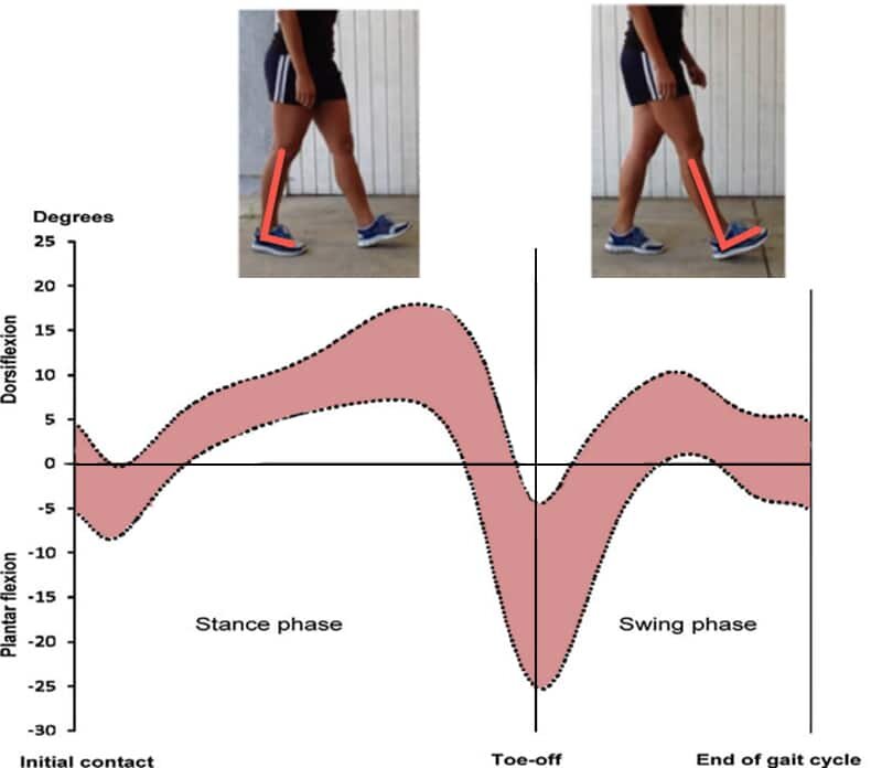
Walking has been shown to require approximately 10 degrees of ankle dorsiflexion by Dr. Jacqueline Perry in her gait research.  As the body advances, the trail leg needs to dorsiflex as the hip extends right before push off.


More recent research has reported that up to 20 degrees of dorsiflexion has been shown in subjects during gait analysis.  

More dynamic movements, such as the squat, require even more ankle dorsiflexion range of motion.  A 2006 study by Hemmerich in the J Orthop Res showed that the further you go into the squat position, the more dorsiflexion motion is needed.  Maximal squat depth to just past parallel requires almost 35 degrees of dorsiflexion.

ankle-dorsiflexion-during-the-squat-845x258.jpg

As you can see in the images above, once you get deeper than 20-30 degrees of squat depth, the amount of dorsiflexion needed goes up considerably. The first image is depicting someone with a restriction with ankle dorsiflexion, and how that will impact the depth of the squat. In the second image, you can see how having more dorsiflexion will improve the depth of the squat. The third photo shows an example of modifying the squat by elevating the heels. This take some of the dorsiflexion needs out of the squat to allow her to go deeper.

Check out this video demonstration on how to improve the squat with limited ankle dorsiflexion to see how we would coach this:

Ever wonder why weightlifting shows have a large heel lift?  Well, it’s to squat deeper by taking out the need for so much dorsiflexion.  In my experience, a lack of dorsiflexion is one of the biggest reasons why people often squat poorly.

How to Assess Your Ankle Mobility

So based on everything we’ve covered so far, you can see, dorsiflexion range of motion can be variable.  That’s why when assessing ankle dorsiflexion mobility, I like to measure the specific degree, but also include a simple self-assessment that people can do themselves.  This is simply a pass-fail type test.

I’m also a big fan of standardizing a test that can provide reliable results.

One test that is popular and part of our integrated movement assessment in the Champion Performance Specialist program is the half-kneeling dorsiflexion test.   Here’s a video from the program demonstrating:

In this test, you kneel on the ground and assume a position similar to stretching your hip flexors, with your knee on the floor.  Your lead foot that you are testing should be lined up 5″ from the wall.  This is important and the key to standardizing the test.

From this position you lean in, keeping your heel on the ground.  You can measure the actual tibial angle in relationship to the ground with a goniometer, or simply measure the distance of the knee cap from the wall when the heel starts to come up.  

An alternate method would be to perform the same movement and measure how many inches forward your knee passes your toes.  If I am looking to quantify this movement to be able to assess progress, I’ll use this method.

I personally prefer to standardize the distance to 5″ from the wall and simply perform it as a pass-fail test.  If they can touch the wall from 5″, they have pretty good mobility.  

I should note that my photo below has my client wearing minimal heel drop shoes, but barefoot is ideal

This is a great position to assess your progress, and as you’ll see, I’ll recommend some specific drills you can perform from this position so you can immediately assess and reassess.


Ankle Mobility Exercises to Improve Dorsiflexion

There are many great ideas on the internet on how to improve dorsiflexion with ankle mobility exercise, but I wanted to accumulate some of my favorites in one place.  Below, I will share my system for assessing ankle mobility and then addressing limitations.  I use a combined approach including self-myofascial exercises, stretching, and ankle mobility drills.

As I mentioned previously, I like to use a 3-step process to maximize my gains when trying to enhance ankle dorsiflexion:

  1. Self-myofascial release for the calf and plantar fascia

  2. Stretching of the calf

  3. Ankle mobility drills

I prefer this order to neuromodulate tone, loosen the soft tissue and maximize pliability before working on specific ankle joint mobility.  

When looking at the efficacy, a recent study showed that combining self-myofascial release and static stretching had a greater increase in ankle dorsiflexion range of motion than either on their own.

Self Myofascial Drills for Ankle Dorsiflexion Mobility

One of the more simple self-myofascial release techniques for ankle mobility is foam rolling the calf.  

Anytime I perform self-myofascial release exercises, I follow a 3-step plan:

  1. Roll up and down the entire length of the muscle for ~10 reps or up to 30 seconds

  2. If they hit a tender spot or trigger point, pause at the spot for ~8-10 seconds

  3. Add active ankle range of motion movements during rolling, such as actively dorsiflexing the foot or performing ankle circles:

A foam roller does a really good job of helping to improve ankle dorsiflexion.  This has benefits as you can turn your body side to side and get the medial and lateral aspect of your calf along the full length.  

Here’s a clip of someone performing this in real-time.  Notice how they hit all aspects of the calf follow my 3-step self-myofascial release system.  Don’t forget to roll the bottom of your foot with a ball, as well, to lengthen the posterior chain tissue even further.  There is a direct connection between the plantar fascia and Achilles tendon.

Some people do not feel that the foam roller gives them enough of a release as it is hard to place a lot of bodyweight through the foam roller in this position.  That is why I often use one of the massage sticks to work the area in addition.  You can use a massage stick in a similar fashion to roll the length of the area and pause at tender spots.  I often add mobility in the half-kneeling position as well, which gives this technique an added bonus.


Stretches for Ankle Dorsiflexion Mobility

Once you are done foam rolling, I like to stretch the muscle.  A systematic review by Radford has shown that static ankle dorsiflexion stretching can lead to a statistically significant improvement in motion.  A more recent meta-analysis revealed a 5-degree increase in mobility after ankle dorsiflexion stretches.

If moderate to severe restrictions exist, I will hold the stretch for about 30 seconds, but often just do a few reps of 10 seconds for most people.  

The classic wall lean stretch is shown below.  This is a decent basic exercise, however, I have found that you need to be pretty tight to get a decent stretch in this position.

I usually prefer placing your foot up on a wall or step instead, as seen in the second part of my video below.  The added benefit here is that you can control the intensity of the stretch by how close you are to the wall and how much you lean your body in.  I also like that it extends my toes, which gives a stretch of the plantar fascia as well.  For both of these stretches, be sure to not turn your foot outward.  You should be neutral to point your toe in slightly (no more than an hour on a clock).


Simple Ankle Mobility Exercises

I like to break down my ankle mobility exercises into basic and advanced, depending on the extent of your motion restriction.  There are several basic drills that you can incorporate into your movement prep or corrective exercise strategies.

Half-Kneeling Dorsiflexion Wall Mobilization

The first drill involves a simple half-kneeling dorsiflexion movement, which is essentially just a dynamic warmup version of the ankle mobility test we described above:

This tends to be the first dorsiflexion mobility exercises that I tend to give people, as it is super easy to perform, and also easy to gauge progress by your distance from the wall.

Half-Kneeling Ankle Dorsiflexion with a Dowel

A progression from the wall mobilization is to use a dowel.  The dowel is an important part of the ankle mobility drill.  You begin by half kneeling, then placing a dowel in front of your 2nd or 3rd toe.  Now, when you lean into dorsiflexion, make sure your knee goes outside of the dowel.  This will help maintain a neutral arch position and avoid compensating by pronating your foot and internally rotating your hip.  You can add the dowel to many of the variations of drills we are discussing.

Half-Kneeling Ankle Dorsiflexion with Your Hand

A slightly more advanced variation of the dowel is to use your hand.  Your arm will act like the dowel, and require your knee to go to the outside.  This movement requires more hip mobility to perform but can be really useful for those that may be struggling to keep a neutral arch during the movement.

Half-Kneeling Lateral Ankle Glides

For some people, especially those with a past injury like an ankle sprain, lateral mobility of the ankle can become limited.  This mobility drill is very popular with my clients, they really like the feeling and ability to move afterward/


Advanced Ankle Mobility Exercises

When the person is ready to progress to more advanced ankle mobility drills, I often like to progress the drills by including multiple planes of motion, making the positions more dynamic, or progressing to drills that require more dorsiflexion to accomplish.

Half-Kneeling Dorsiflexion with Voodoo Floss

Chris Johnson shared a nice video using a Voodoo Floss band to assist with the myofascial release and position the tibia into internal rotation.  Restoring ankle dorsiflexion at the level of the talocrural joint is critical and should be established with the knee flexed to mimic the midstance demands of walking and running. The performer will grasp the distal tibia and fibula region and wind into internal or external rotation and then lunge forward and backward. 

Half-Kneeling Dorsiflexion with a Mobility Band

For those that have a “pinch” in the front of the ankle of tight joint restrictions of the ankle in general, Erson Religioso shows us some Mulligan mobilizations with movement (MWM) using a band.  In this video, he has his patient put the band under his opposite knee, however you could easily tie this around something behind you.  In this position you step out to create tension on the band, which will move your talus posteriorly as you move forward into dorsiflexion:

Standing Dorsiflexion Wall Mobilization

The standing dorsiflexion wall mobilization is an advanced version of the half-kneeling position.  But rather than simply leaning in and touching your knee to the wall, we change the start position to that your toes up on the wall and extended.  This essentially combines an ankle mobility drill with a stretch to the calf, Achilles tendon, and even the plantar fascia due to the toes being extended.

This isn’t one of the first drills I use because it’s advanced, but it’s one of my favorites.

Standing 3-Way Dorsiflexion Wall Mobilization

Kevin Neeld shows a great progression of this exercise that incorporates both the toes up on the wall, essentially making it more of a mobility challenge and stretch.  If you look closely, you’ll see that he is also mobilizing in three planes, straight neutral, inward, and outward:

Standing Lateral Ankle Glides

Similar to the above progression from half-kneeling to standing dorsiflexion drills, I like to perform a more advanced version of the lateral ankle glides by performing standing:

Push Up Position Ankle Rocks

As you can see, we’re really progressing to positions that require more and more ankle dorsiflexion range of motion, so you can see the potential progressions.

The push up position ankle rocks drill is a great one, that involves a great stretch of the calf and Achilles tendon.  Most of my athletes will use this ankle dynamic mobility drill at some point in their warm-up.


Functional Dorsiflexion Dynamic Mobility Drills

So far, we’ve covered a variety of mobility drills to perform to enhance ankle dorsiflexion.  As mobility increases, it’s super important that we try to functionally incorporate dorsiflexion into our programming.

Here are some examples that I like to use.

Seated Dorsiflexion Ankle Raises

Once you’ve established dorsiflexion mobility, it’s a good idea to strengthen the muscles that dorsiflex within the new range of motion.  A nice easy drill to start with is seated dorsiflexion ankle raises.  Because you start in the neutral position, any lift of the ground is moving towards more dorsiflexion

Standing Deficit Dorsiflexion Toe Raises

A variation of the seated position, standing up on a weight plate or similar low height box will allow you to need to dorsiflex against any tightness of the gastrocnemius.  By adding the box, you will allow strengthening into a great range of motion.

Knee Extension with Dorsiflexion

In addition to strengthening into dorsiflexion, I also like to work on the endurance of maintaining the position.  An easy way to do this is with a kettlebell around the toes of the foot.  You will need to dorsiflex to hold the kettlebell, and can easily progress to heavier weights.  I often incorporate this with a knee extension movement to include a lengthening of the gastrocnemius.

Toes Elevated Squats

As we’ve previously discussed, the squatting motion requires a lot of dorsiflexion to go deep into the motion.  

One drill I like to do for a dynamic warm-up prior to squatting in someone that is working to increase their dorsiflexion is simple toes elevated squats.  You can slide a small weight plate, or something about 1-1.5”, under the toes and slowly perform some bodyweight squats.

Reverse Bear Crawls

The reverse bear crawl is an amazing exercise for ankle dorsiflexion.  When crawling backward, you’ll need a decent amount of ankle mobility.  The movement includes a dynamic movement into dorsiflexion with the knee bent and toes extended, so acts as a good drill for the soleus and plantar fascia.

Improving Dorsiflexion Mobility

As you can see, there are many different variations of drills you can perform based on what is specifically tight or limited.  You may have to play around a little bit to find what works best for each person. However, these are a bunch of great examples of ankle mobility exercises you can choose to perform when trying to improve your dorsiflexion.

Think you have poor posture? Maybe it's not what you think..........

AdobeStock_186427438.jpeg

Why “Stand Up Straight” Was Never a Proper Instruction: Posture Getting Worse?

Head up, shoulders back, pain solved. That’s how it’s supposed to go. But what if everything you’ve been told about “good posture” is wrong, and in fact, is only making your pain worse?

This is the question I’ve been asking myself for months now, ever since — after almost a decade of nagging left shoulder pain that wouldn’t go away — a practitioner I found on YouTube finally healed me.

Seeking medical advice on YouTube? I know, I know. But I was at the end of my rope with my body — and my doctors. For years, every expert I consulted told me the same thing: “You hunch over a computer all day. Of course your shoulder hurts.” One doctor told me the problem was my pathetic upper body strength, so I got really into power yoga. I practiced my posture during my commute by closing my eyes and remembering the mantras I learned in class: “Lengthen the spine” and “squeeze the shoulder blades.” I got massages once a month. But alas, no matter how well I could propel myself from downward dog to chaturanga, no matter how much length I gave my spine or how much I was kneaded, when I looked in the mirror, I saw the same slouchy woman. And my shoulder still ached.

Good posture has always been more of a cultural ideal than anything else, “a litmus test for the healthy, modern body of the perfect citizen.”

Most of us take standing up straight for granted as basic health advice. We’re berated about it by our parents and teachers as tweens, and as adults in a capitalist society, we’re bombarded with the same messaging buried in the promises of proper ergonomics, standing desks, and “smart devices” that will ping your phone if you start to slouch. But as I learned the hard way, very little of this has anything to do with actual health or science.

How to Ease Your Quarantine Aches and Pains

Here’s why your body hurts and what you can do about it

Good posture has always been more of a cultural ideal than anything else, “a litmus test for the healthy, modern body of the perfect citizen,” as Emory University professor and cultural historian Sander Gilman, PhD, writes in his 2018 book, Stand Up Straight: A History of Posture. The concept of “good” or “bad” posture dates back to ancient times: Hippocrates himself wrote about poor habits being a possible cause of spinal malformation. But to understand why he thought that, you have to know that the ancient Greeks understood posture as the defining character of mankind, Gilman says. Greek myths are rife with postural lessons. There’s Thersites, the hump-backed and dull-witted soldier who gets beat up by Odysseus for causing trouble during the Trojan Wars, and posture and gait is even a recurring theme in the tale of Oedipus. The Greeks believed that man stood upright to face the Gods.

This cultural understanding has followed us ever since, from the 19th-century rise of Swedish gymnastics to the bizarre “Miss Correct Posture” pageants of the 1950s, to the wearable “smart” devices of today. “We have this unspoken language of posture: It affects the way we see other people,” Gilman says. “It affects the way we see our own selves in the world.”

As ridiculous as some of these examples seem now, our assumptions of beautiful posture, or even just an “appropriate” posture, infect our ideas about what makes a “healthy” posture as well. Today, back pain is one of the most common health complaints. “Something like 80% of people will experience back pain at some point in their lives, and it can be debilitating,” explains Charles Kim, MD, a physiatrist at NYU Langone Health. Posture remains among the first things many doctors and physical therapists ask about if you report back pain, despite the fact that multiple recent studies suggest the link between back pain and posture is weak, if not nonexistent. Take this 2016 metanalysis of 10 studies that couldn’t confirm a link between “excessive thoracic kyphosis” (the technical term for slouching) and shoulder pain. Or this 2018 study that found no link between “text neck” (the technical term for looking down at your iPhone) and neck pain in 624 young adults.

This isn’t to say that slouching or looking down at your phone won’t ever lead to pain. Sitting or standing in any one position repeatedly can cause certain stresses on your joints over time. But this idea that there is singularly one healthy posture for everybody — regardless of differences in size or asymmetry or what daily activities you do — is what I’ve found to be dangerously bunk.

It wasn’t until my left knee also started hurting, making it impossible to run, that the “good” posture myth finally fell apart for me. The discovery was all thanks to a YouTube channel run by Neal Hallinan, a certified strength and conditioning specialist, called “The PRI Trainer.” PRI stands for Postural Restoration Institute, a growing school of thought in physical therapy. In the past 30 years, the institute, led by Ron Hruska, a doctor of physical therapy (DPT), has trained more than 200 physical therapists, athletic trainers, and even dentists in helping people correct what they see as the real troublemaker: poor breathing mechanics.

“When people get in that tall, erect position, they do not have their diaphragm in a position to work appropriately as a respiratory muscle.”

The basic idea behind PRI’s teachings is that human bodies are naturally asymmetrical, and this affects how we move and breathe. Although you are likely pretty symmetrical on the outside (two arms, two legs, two breasts, etc.), internally, your heart sits on your left side, and to make room for it, your left lung is slightly smaller than your right lung. Your main breathing muscle, the diaphragm, is actually two muscles, a stronger, larger right leaflet and a smaller, weaker left leaflet. “Because of this, the left and the right side are not doing the same thing,” says Jennifer Platt, DPT, executive director of the Postural Restoration Institute.

Problems begin when stress and repetitive activities in our lives accentuate this natural imbalance. “Walking, riding the bike, or sitting at the computer, doing any of these repetitive activities, you become very lateralized. You become one-side dominant,” Platt says. And that can wreak havoc on your neck, shoulders, back, and joints. Even worse: The advice to lengthen the spine can aggravate things. “When people get in that tall, erect position, they do not have their diaphragm in a position to work appropriately as a respiratory muscle,” Platt says. “That reinforces the pattern.”

What I learned from PRI training, which consists of targeted breathing and isolation techniques, is that my posture really was terrible — but not in the traditional “up and down” sense. Rather, my posture was terrible because I was tragically uneven left to right. Why didn’t any of the number of professionals I consulted about my pain ever mention this? “Back pain especially, but pain in general, is very poorly taught in traditional medical education,” Kim says.

Physical therapists aren’t always much help, either. “In PT school, you learn the anatomy, but you don’t really learn the whys,” Platt says. “The connection hasn’t been made between the internal asymmetry and the function of the human body.”

Reading the reviews on Neal Hallinan’s website makes PRI seem way too good to be true. “I used to suffer pain literally 24/7,” one reads. “One night I woke up in pain at 2 a.m., found your exercises, and when I did them I felt so good that I finally had a deep sleep.” Normally, I’m a skeptic of miracle fixes. But when I reached out to Hallinan in January of this year, I was ready to try anything.

A few months prior, I had run my first marathon. It didn’t go that well — both my left knee and my left shoulder had locked around 10 miles in, stealing my time goal and twisting me into a pretzel for my finish line photos. After the race, my body simply had it. My shoulder hurt more than ever, and I was diagnosed with IT band syndrome in the left knee and tendonitis in my right Achilles.

Worse, I couldn’t seem to recover no matter how much I rested. After months of physical therapy that didn’t help, I paid $300 for a gait analysis that revealed my left hip didn’t load while I ran. This seemed to explain at least some of my issues: Hip weakness, aha! What I thought I’d get from Hallinan was an exercise program to strengthen that hip. I did not expect him to tell me during our first virtual session that I wasn’t struggling with “weakness” necessarily, but with an inability to connect to the appropriate muscles to actually use my left hip. Even less did I expect him to say that the real issue — the true source of all of my problems — was that I was bad at breathing.

It’s hard to be told you don’t know how to breathe. (Isn’t it, you know, automatic?) But if PRI founder Ron Hruska’s theories are correct, a lot of us are breathing inefficiently. “Most people are breathing with too much compensatory muscle because of the position that they are stuck in,” Hallinan says.

The diaphragm is the main breathing muscle. But we also have accessory breathing muscles in between our ribs, called the external intercostals, as well as certain muscles in the neck. Your diaphragm on both sides contracts as you inhale, pulling down and creating space in your chest for the lungs to expand. It’s like a plunger that pulls air into your lungs. At the same time, your intercostals pull your ribs up and out, creating even more space for air to fill the lungs. During exhalation, everything relaxes.

The key to all of this working is making sure your diaphragm remains in the domed position so it can plunge efficiently. The area where the dome of the diaphragm sits is called the “zone of apposition” or ZOA, and it is heavily influenced by the orientation of the rib cage. If the ribs are off, you can shift into a position where the dome is lost. Maintaining the dome not only helps you breathe efficiently, but the plunging action also helps keep your pelvis, spine, and ribs stable because it, along with the pelvic floor, creates intra-abdominal pressure, explains Leada Malek, DPT, a board-certified sports physical therapist based in San Francisco.

But remember: That left leaflet of the diaphragm isn’t quite as big as the right one. “It’s the weak link, our true Achilles’ heel,” Hallinan says. So what happens is we can very easily lose the dome shape on the left side, which means it can’t create that plunging action. The right diaphragm, intercostals, and sometimes the right-side neck muscles then take over to make sure we can get air into our lungs.

“This can breed compensatory and dysfunctional movement patterns,” Malek says. “A ‘chest breather,’ for example, will move while using accessory muscles of breathing that attach to the upper ribs and can contribute to significant tightness around the neck. This can then cause movement dysfunctions in the shoulder.” If things continue, a cascade of issues can crop up from there.

For me, it started with shoulder pain and escalated to my left knee and right foot. But where pain shows up doesn’t always follow a strict pattern. “I always say the body is so smart. It’s too smart sometimes,” Malek says. “Your body will find one way or another to stabilize you, and over time, that leads to stress somewhere.”

For Cynthia Cook, a personal trainer in New York City, it manifested as dreaded low back pain, a relentless tightness that got so bad on her right side she couldn’t walk for two months. “One would say I have perfect posture. I was doing barre classes. I was in the military,” she says. As a fitness professional, she’s also hella strong. But none of that stopped her from experiencing debilitating low back pain. X-rays revealed nothing going wrong with her spine, and traditional physical therapy didn’t help much, either.

“I was in really severe pain and nothing worked. It never made sense to me that I just needed to strengthen something or stretch something,” she says. Eventually, like me, she found PRI through Google and the rest is history. “I’ve never felt better.”

For the past eight months, I’ve started every day with at least 15 minutes of PRI training. There are a number of different techniques that PRI trainers use based on what’s going on with a person’s body — or how deeply stuck in a certain movement pattern they are. But the key to all PRI exercises is that you exhale slowly, making sure you push all the air out. (This helps you reposition your ribs.) In normal breathing, exhalation requires no effort. But elongating the exhale is itself a kind of workout that activates the internal obliques. Making these specific abdominal muscles stronger helps you keep your ribs on the left side down and your diaphragm and ribs in the optimal position, even during normal breathing.

A good example is the 90–90 hip shift, which is one of the more common techniques in PRI. To do it, you lay on your back on the floor with your feet resting on a wall, so your knees create a 90-degree angle. You place a ball or towel in between your knees and squeeze. Then, you lift your butt slightly off the ground so your pelvis is tilted and your feet are pressing into the wall. Then, you shift your right knee slightly toward the ceiling and begin to lift your right foot so your weight fully shifts to the left side. Finally, while you’re doing all of that, you breathe deeply, trying to make your exhales twice as long as your inhales.

There’s a lot going on here—but that’s because you can’t separate learning to breathe efficiently from learning to move efficiently. “When you try to think about A leading to B leading to C you get into trouble,” Hallinan says. “Everything occurs at once.”

Thanks to consistent practice with these kinds of exercises, I feel amazing — and I’m slouchier than ever. Whenever my shoulder acts up, I know how to take a few breaths and get into the right position to turn off the tension. And what you’ll find doing PRI is that you pretty much have to relax your shoulders to do these techniques correctly.

I look in the mirror today and have a new appreciation for the fact that I slouch, and that my body isn’t exactly symmetrical.

Of course, it’s important to point out that breathing inefficiency isn’t the only thing that causes back pain. Injuries, spinal issues, arthritis, pulled muscles — all of those things can make you hurt, Kim says. But even he admits that something like 60% of people with back pain have no spinal abnormalities or injuries. Could the reason those people’s backs hurt be a breathing issue? Kim puts it at maybe. From his perspective, PRI is an interesting set of theories.

Hallinan and Platt put it at most likely. Malek says she uses PRI in her practice any time a client just isn’t getting better with the regular techniques. “[Posture] is the simple answer. Sometimes sitting up straighter is all you need, but if you keep chasing it and it’s not helping, then what do you do?” she says.

At the end of the day, it’s hard to say who’s right. The natural asymmetry of the diaphragm is well documented in scientific literature, and a growingbody of research supports the use of PRI techniques for a variety of frustratingly common pain syndromes, like IT band syndrome and low back pain. But the studies tend to be small. The good news is PRI training is not going to hurt you. And if you’re in pain, anything that won’t hurt you more can be worth a try, Kim says.

Of course I’m glad I found a way to solve my pain. But the best part is that I gained a deeper awareness and acceptance of my body as it is. I look in the mirror today and have a new appreciation for the fact that I slouch, and that my body isn’t exactly symmetrical. Instead of forcing my spine straight and my shoulders back, I revel in the fact that I can move and breathe in this perfectly imperfect body.

The more I look back on how long I spent in pain, the more I realize that I really did believe my pain was the result of some moral failing, like if only I wasn’t so lazy or weak, I’d have learned to straighten it out (literally). “One of the greatest difficulties we have is that posture like many other qualities of the body is also associated with character,” Gilman says.

If this were ancient Greece, I would not be considered a model citizen — and I’m okay with that.

Amelia Harnish

Health and culture writer based in Ridgewood, Queens. Slow runner. ameliaharnish.com


More information from the Postural Restoration team at Pinnacle PT: PRI at Pinnacle PT

5 Muscles You Think You Are Training

From our friends and mentors at the Postural Restoration Institute and The Hruska PT Clinic…..

5 MUSCLES YOU THINK YOU'RE TRAINING... BUT YOU'RE NOT!

What to do when good treatment just doesn't work

If you have seen a Physical Therapist for a rehabilitation program or done any type of strength and conditioning program to improve your strength or performance, with or without the help of a certified trainer, you have been instructed in a program that aims to train specific muscle groups. This is the purpose of rehabilitation or training.

But, what if you are doing everything in that rehab or performance program you should and are just not seeing the results you want. Maybe your back still hurts after weeks or months of “core” strengthening exercises you got from the PT. Or maybe you just haven’t gotten that much faster or stronger in your jumping despite having your butt kicked with all that glute strengthening activity you’ve been doing for months. What’s that all about?

Here’s the deal. Many trainers and Physical Therapists have great knowledge in what needs to get stronger and can give a great program to strengthen specific things to get the results you should get.

They are just missing one really important concept.

Muscles have specific alignment needs in order to be able to work effectively. The position you place a muscle in has a significant impact on how well it can actually contract and do the things you are asking it to do. If it can’t effectively work the way you are asking it to, your brain will automatically ask other muscles, that aren’t as good at the task you are trying to do, to “help out”. This effectively allows the muscles that you are training to stay weaker while at the same time may be allowing other undesirable muscles to get stronger.

Muscles are placed in a specific alignment by the position, or posture, they are placed in either purposely or unconsciously when you are training them. At the Hruska Clinic we understand normal patterns of human movement and posture that renders some muscle groups ineffective at doing their job. Unless this is addressed first any rehab or training program, even programs with the correct activities, will be ineffective at best, and potentially could be actually making you feel worse by doing them.

Here are commonly trained muscle groups that are often not accessible for effective training unless muscle position and posture is addressed first.

1. Glutes

Your gluteal muscles, or glutes, are actually a combination of several muscle groups that attach from your pelvis to your thigh bone at the back and side of your hip. You actually have three glutes on each side (along with some other smaller deep hip muscles)! You have the well known Gluteus Maximus which is a strong outer rotator of the thigh and extender or the thigh. It is the largest of the gluteal group and is your primary pusher (or should be) when doing things like stairs or squats or

lunges. You also have your Gluteus Medius and Gluteus Minimus which are underneath the Gluteus Maximus and are more responsible for turning the thigh bone in and taking the thigh bone out to the side. Their primary role functionally is to stabilize the hip when you are standing on one leg. When you are balancing on one foot (which only happens every step you take) this glute group keeps the hip joint stable so you can balance and push yourself to the next step with your “push” glutes.

AdobeStock_156731686.jpeg

These muscles are very
commonly trained and isolated
in rehab programs for general
leg strength like after any leg
surgery or injury. They are
needed for any activity done in standing so if you are having pain or issues with any activity in standing your glutes will probably be trained. For performance purposes glutes are necessary to gain more power and control with running, jumping and other sport specific actions.

Both of these glute groups are strongly influenced in their ability to do their jobs based on the position of the pelvis and the thigh bone. The pattern or position in which you hold yourself up when you stand or walk will have a direct impact on the position of these muscle groups which in turn influences how well or effectively they can do their job. If this position is not ideal, and not addressed, you will compensate to push or stabilize with other muscle groups that may not be as good at that job as your glutes.

For example, if you are doing and activity that requires pushing with your gluteus maximus like a squat, lunge or a ‘glute bridge’ and that muscle can’t work as effectively as you’d like, you will engage some of your other "pushers". If you’ve gotten done with a “glute” workout and your back is sore, or your knee is sore or hurts during those activities, you may not have access to your gluteus maximus muscle the way you’d like. If you are doing a stabilizing activity, or activity on one leg (walking and running included) and you can’t access your stabilizer glutes you will engage other less effective stabilizers. This undesirable compensation can lead to potential issues with your thigh, knee, back or deep hip joint.

What we understand at the Hruska Clinic, based on science taught through the Postural Restoration Institute , is that our body is biased for more stability on the right leg and more push on the left leg. This is based on postural and functional asymmetries that are common human characteristics. What this means for your glutes is that you will have better access to your right hip stabilizers (Medius and Minimus) and better access to your left pushers (Maximus). Unless this imbalance is addressed, glute programs may not give you the desired results you want or may be creating pain patterns in areas you don’t want.

2. Abdominals

When someone is started on a “core” strengthening program the abdominal group is the main (and sometimes only) muscle group people think of. This group of muscles attaches to your pelvis bone and your sternum and rib cage in the front and sides and in the case of the deep layer of muscles runs from one side of your lower back/spine all the way around to the other side like an internal belt or brace. It’s primary job (other than looking good on the beach) is to stabilize your abdominal contents and reduce strain on your lower back as well as to help create movement of your trunk needed for rotation and power along with stabilizing the midsection of your body so your extremities can function to do their jobs effectively.

They are used ALL the time when you are upright and moving. This is why they are, and should be, a part of any strength and conditioning program and will be involved in just about every lower back rehabilitation program I’ve ever seen. Because of their attachment on the pelvis bone and ribs their ability to function correctly is directly influenced by the position or posture of the pelvis and rib cage. An anteriorly tipped pelvis will lengthen the abdominal muscles and render them less efficient. “Flared” or outer rotated ribs will also limit abdominal efficiency.

If your abdominal muscles are not in the correct position when you are training them you will utilize other muscle groups to provide that “core” stability you require. Commonly over trained “core” muscles that will engage for your abdominals are your hip flexors, which can stabilize your spine and hip in an anteriorly tilted pelvis position, your lower back muscles which will also work harder to stabilize and protect your spine if your abs aren’t helping out, and your diaphragm or breathing muscle that will try to stabilize your spine, ribs, and abdominal contents if the ab group is failing at that. If you get done with a “core exercise” and have front hip pain, lower back pain or can’t breathe well, during or after an activity, you may be compensating and not utilizing your abs well.

Therefore, the position of both the pelvis and rib-cage will need to be addressed prior to and during any abdominal activity. Both of these areas, your pelvis and rib cage, are positioned in a pattern for upright posture by a neurological process in your brain. There are many factors taken into consideration by your brain to ‘set’ this postural pattern, like the surface you are standing on, the activity going on around you and your current strength or fatigue levels. If your upright pattern or posture is imbalanced, this WILL directly affect your abdominal efficiency. We understand those patterns of activity and the myriad of things that may be influencing them, and will address them to ensure you actually develop an effective core for performance and injury rehabilitation.

3. Lower Traps

AdobeStock_303415062 (1).jpeg

The lower trapezius, or lower trap, muscle is a muscle that lies on the back of your shoulder blade. It runs from the main spine
of your shoulder blade down and in to connect to many of the thoracic vertebrae on your back. It is a muscle that pulls your shoulders “down and back”. This is a common cue for many programs and exercises to stabilize the shoulder blade, improve forward shoulder posture and to protect your shoulder, or rotator cuff, when doing arm activities. If the lower trap muscle isn't working well the shoulder blade, which is the socket for your shoulder joint, will not be able to allow the muscles of the shoulder and arm to work well. For this reason this muscle is isolated and trained not only for posture, but also for overhead athletes to prevent arm injury, improve arm function for throwing, and is a key scapular stabilizer in shoulder rehab in surgical and non-surgical cases.

If the lower trap isn’t stabilizing the shoulder blade you may develop compensatory muscle activation of the upper trap (neck), latissimus dorsi (lower back), posterior rotator cuff, among others. Another often overlooked impact of the weakness or imbalance between the lower trap muscles is in their ability to stabilize and rotate the middle thoracic spine.

In order to effectively train the lower trap the position of its attachments, the shoulder blade and thoracic spine, must be addressed to ensure proper muscle alignment. The shoulder blade is a large bone that sits on top of the rib
cage. It is not actually attached to the rib cage except through muscle attachment. Therefore the position of the shoulder blade is directly impacted by the position of the ribs underneath it. The thoracic spine moves primarily in the forward and backward or rotational directions. The rotation of the thoracic spine is also directly influenced by, and influences, the position of the rib cage.

Due to this anatomy the primary factor that needs to be addressed to ensure the lower trapezius is in a position to function well is the position of the rib cage and thoracic spine, not just the shoulder itself.

Unfortunately, it too often is not.

Your rib cage moves (expands and contracts) as you breathe. Therefore, breathing and how you breathe with proper rib mechanics directly affects rib position, spine position, scapular position and ultimately,lower trap position. Breathing mechanics along with spine position are too often missed in shoudler rehab or performance training.

If you haven’t gotten the results you want from your shoulder performance or rehab program and no one has looked at your breathing, perhaps that needs to be addressed so your program can actually work.

4. Pelvic Floor

The pelvic floor is actually a group of muscles that are attached to the base of the pelvis and work somewhat like a sling or hammock to support your internal organs as well as giving support to the pelvis bones themselves. There are three main bones that make up the pelvis itself. The sacrum and the right and left innominate bone. The sacrum is the wedge shaped bone at the base of your spine that extends into your tailbone (or coccyx) and the innominates are the “hip” portion of your pelvis that houses your hip socket. That bone is actually also divided up into 3 sections (the ilium, the ischium and the pubic bones.) The pelvic floor muscles are named for which of these bones they attach to. (I.e the pubococcygeus attaches the pubic bone to the coccyx) Functionally it is hard to isolate one pelvic floor muscle from another which is why we group them as your pelvic floor muscles.

AdobeStock_317008354 (1).jpeg

The most common reason that someone may be training the pelvic floor muscles is for what is termed pelvic floor dysfunction (PFD). Pelvic floor dysfunction is associated primarily with incontinence but can be also related with other pelvic pain issues. The gold standard exercises for pelvic floor dysfunction are Kegel exercises which are just exercises to intentionally contract your pelvic floor muscles to strengthen them. Biofeedback units are also often used to train people to ensure those muscles are contracting well. Unfortunately some people just don't seem to make progress despite doing Kegel exercises literally for years!

When Kegel exercises aren’t working as well as we would like what has happened, as with our other challenging muscles, is that the position of the muscles wasn’t addressed prior to starting or performing this program. Because the pelvic floor muscles are attached to the pelvis bones their ability to function in the manner they should is directly associated with the position of the pelvis bones. Anything that affects the position of the pelvis bones has an ability to influence the position, and therefore function, of the pelvic floor muscles. Pelvis bone position is influenced by many things but the things we specifically would address are your postural patterns when standing, your ability or inability to shift your center of gravity from side to side with walking, and your foot mechanics with walking (yes your shoes can have a major impact on your pelvic floor function!) among others.

Another key factor and purpose of the pelvic floor is its role in proper diaphragmatic breathing. Your ability or inability to breathe with a normal diaphragmatic breathing pattern is of primary importance to ensuring correct pelvic floor function. Once we address issues that impact your pelvis position and breathing patterns, the ability for the pelvic

floor to function correctly is dramatically improved. If your Kegels aren’t working, perhaps we should look
at some other things.

5. Hamstrings

One of the most often trained or treated muscles by both Physical Therapists and trainers is the hamstring muscle. This muscle, located on the back of the thigh, is stretched, pounded on, scraped, beat up and can be the bane of existence for most people. The hamstring is actually a group of 3 muscles that attach on the base of the pelvis bone, and the back of the thigh bone and run down to the shin bones. Its primary role is often thought to be bending or flexing your knee, which it definitely does, but it is also a very strongly influential muscle for

control of forward and backward tipping of the pelvis. Tightness, or perceived tightness, in the hamstring on one side or both is often seen with people who have lower back pain, hip pain, and front knee pain. Therefore in order to “treat” those issues hamstring flexibility is often a main goal of programs. Let the stretching begin!

However, this hamstring tension that is felt and seen is often more of a product of the position of the hamstring and the demand placed on it rather than the true length of the hamstring itself. In many people, especially those with lower back pain and tightness, the

AdobeStock_267942231 (1).jpeg

pelvis is tipped anteriorly. This position of the

pelvis effectively moves the attachment points of the hamstring (the

knee and the base of pelvis) further away from each other

which actually puts the hamstring muscle into a tighter position before we even start to address it. Whether this happens on one side or both this tilt also increases the demand and need for tension of that muscle as its job is to try and control the pelvis bones. At this point the hamstring is now actually long, overactive and in a poor position. No wonder it feels tight.

Neutral pelvis = Anterior tilt = normal hamstring "tight" hamstring length

This training problem is not that we are addressing the wrong muscle, it is in how we are addressing that muscle. This hamstring does not need to be stretched. In fact stretching it actually places increased demand for tension on the muscle and can have the opposite effect that we are desiring. The position of the hamstring needs to be addressed, especially at the pelvis. If it feels tight it probably actually needs more strength to control the tip of the pelvis, not more stretch. It’s amazing to see a hamstring that only has half of its available motion go to a normal length in seconds, just by getting it a little stronger and in a better position. We do it all the time.

If you’ve been stretching your hamstrings like crazy and they are still “tight” maybe it’s time to try something new.

Conclusion

Nearly every treatment program for rehabilitation or performance addresses and trains specific muscles to try and accomplish their goals. And they should. Unfortunately, for sometimes these well designed programs just don’t give us the desired outcomes whether pain relief or improved performance. Without addressing why certain muscles may not be able to work the way we want them to, even the best programs are doomed to fail.

When you understand some predictable and common patterns of human movement and performance, as well as the factors related to those patterns, you can ensure that the muscles we want to be training can actually be accessed to be trained. Our staff at the Hruska Clinic understands these patterns and everything that influences them, and has the tools available to truly address them. If you have been struggling with these muscles, or others, and your rehab or performance hasn't seen the improvements you would like, give us a call and we will be more than happy to help you access these muscles in a more efficient way.

Visit our website: www.pinnaclephysicaltherapy.org

https://pinnaclephysicaltherapy.org/become-a-patient

For more information from the friends at the Postural Restoration Institute and Hruska PT:

www.hruska-clinic.com Hruska Clinic Restorative Physical Therapy Services 402-467-4545

Fatigue of the Rotator Cuff Causes Altered Shoulder Mechanics and Impingement 

shoulder_impingement_intro01_1.jpg

Fatigue of the Rotator Cuff Causes Altered Shoulder Mechanics and Impingement 

  • Another interesting article from the most recent issue of Journal of Athletic Training on rotator cuff fatigue and glenohumeral kinematics. In this study, the authors used dynamic fluroscopic video to assess superior humeral head migration in 20 asymptomatic subjects before and after fatigue of the rotator cuff. The assessment of migration during a dynamic activity is a fairly novel approach as previous studies that have attempted to quantify superior humeral head translation have used static imaging such as radiographs and MRI.

    During the study the authors used the prone horizontal abduction (with thumbs up) exercise to fatigue the cuff until there was a documented decrease of at least 40% strength. This was an important part of the study, as a reader I want to be sure that fatigue had occurred and a 40% drop is significant for me. I must say, though, that I wish the authors had performed a more specific rotator cuff exercise, such as simple external rotation. The prone exercise has shown EMG activity of the cuff but also the deltoid and scapula musculature.

    Results of the study show that humeral head migration increased by an average of 0.79mm during elevation in a fatigued state. While this seems small, keep in mind that the average subacromial space is between 2mm and 14mm in healthy subjects (can be about 50% smaller in pathological patients), thus reducing subacromial space by up to 40%!

    Clinical Implications

    RTC fatigue can lead to 
    shoulder impingement. An interesting component of the article that the authors did not bring up in their discussion was that the humeral head actually migrated inferior prior to fatigue as the RTC acted to maintain the humeral head within the glenoid fossa as the deltoid provided a superior orientated force vector. After the fatigue, not only did the humeral head not migrate inferior, it moved superiorly. This has obvious implications for subacromial impingement. Both strength and endurance of the cuff should be addressed in rehabilitation, makes sense why a deconditioned person so much more likely to develop shoulder impingement.

    The rotator cuff should never be worked to failure. The fatigue protocol used by the authors lasted for less than 90 seconds. That is all it took for the RTC to loose 54% of it’s strength. Now think about that last time you had a patient come to you with “impingement” that reported playing tennis or golf for the first time in months, or even more simply, painting a room in their house. With only 90 seconds of activity the cuff fatigue enough to decrease your subacromial space by up to 40%. What should your treatment be for that patient? Minimize the initial inflammation, work on cuff strength & endurance but by all means, DON’T work the cuff to failure.

    The rotator cuff should NEVER be worked to failure!

    I never understood this training technique. If you work the cuff to failure, then what is going to dynamically stabilize the humeral head later that day when you are reaching overhead?


Read the full article here:

Exercise as effective as surgery for middle aged patients with knee damage Study raises questions over widespread practice without supporting evidence

AdobeStock_198669548.jpeg

Summary:Exercise therapy is as effective as surgery for middle aged patients with a common type of knee injury known as meniscal tear (damage to the rubbery discs that cushion the knee joint), finds a new study.Share:

FULL STORY

Exercise therapy is as effective as surgery for middle aged patients with a common type of knee injury known as meniscal tear (damage to the rubbery discs that cushion the knee joint), finds a study in The BMJ this week.

The researchers suggest that supervised exercise therapy should be considered as a treatment option for middle aged patients with this type of knee damage.

Every year, an estimated two million people worldwide undergo knee arthroscopy (keyhole surgery to relieve pain and improve movement) at a cost of several billion US dollars. Yet current evidence suggests that arthroscopic knee surgery offers little benefit for most patients.

So researchers based in Denmark and Norway carried out a randomised controlled trial to compare exercise therapy alone with arthroscopic surgery alone in middle aged patients with degenerative meniscal tears.

A randomised controlled trial is one of the best ways for determining whether an intervention actually has the desired effect.

They identified 140 adults (average age 50 years) with degenerative meniscal tears, verified by MRI scan, at two public hospitals and two physiotherapy clinics in Norway. Almost all (96%) participants had no definitive x-ray evidence of osteoarthritis.

Half of the patients received a supervised exercise programme over 12 weeks (2-3 sessions each week) and half received arthroscopic surgery followed by simple daily exercises to perform at home.

Thigh muscle strength was assessed at three months and patient reported knee function was recorded at two years.

No clinically relevant difference was found between the two groups for outcomes such as pain, function in sport and recreation, and knee related quality of life. At three months, muscle strength had improved in the exercise group.

No serious adverse events occurred in either group during the two-year follow-up. Thirteen (19%) of participants in the exercise group crossed over to surgery during the follow-up period, with no additional benefit.

"Supervised exercise therapy showed positive effects over surgery in improving thigh muscle strength, at least in the short term," say the authors. "Our results should encourage clinicians and middle aged patients with degenerative meniscal tear and no radiographic evidence of osteoarthritis to consider supervised structured exercise therapy as a treatment option."

How did this situation -- widespread practice without supporting evidence of even moderate quality -- come about, ask two experts in a linked editorial? "Essentially, good evidence has been widely ignored," say Teppo Järvinen at the University of Helsinki and Gordon Guyatt at McMaster University in Canada.

"In a world of increasing awareness of constrained resources and epidemic medical waste, what we should not do is allow the orthopaedic community, hospital administrators, healthcare providers, and funders to ignore the results of rigorous trials and continue widespread use of procedures for which there has never been compelling evidence," they conclude.

Story Source:

Materials provided by BMJNote: Content may be edited for style and length.

Journal Reference:

  1. Nina Jullum Kise, May Arna Risberg, Silje Stensrud, Jonas Ranstam, Lars Engebretsen, Ewa M Roos. Exercise therapy versus arthroscopic partial meniscectomy for degenerative meniscal tear in middle aged patients: randomised controlled trial with two year follow-up. BMJ, 2016; i3740 DOI: 10.1136/bmj.i3740