Lateral Pelvic Tilt: Its Cause and How to Fix It!
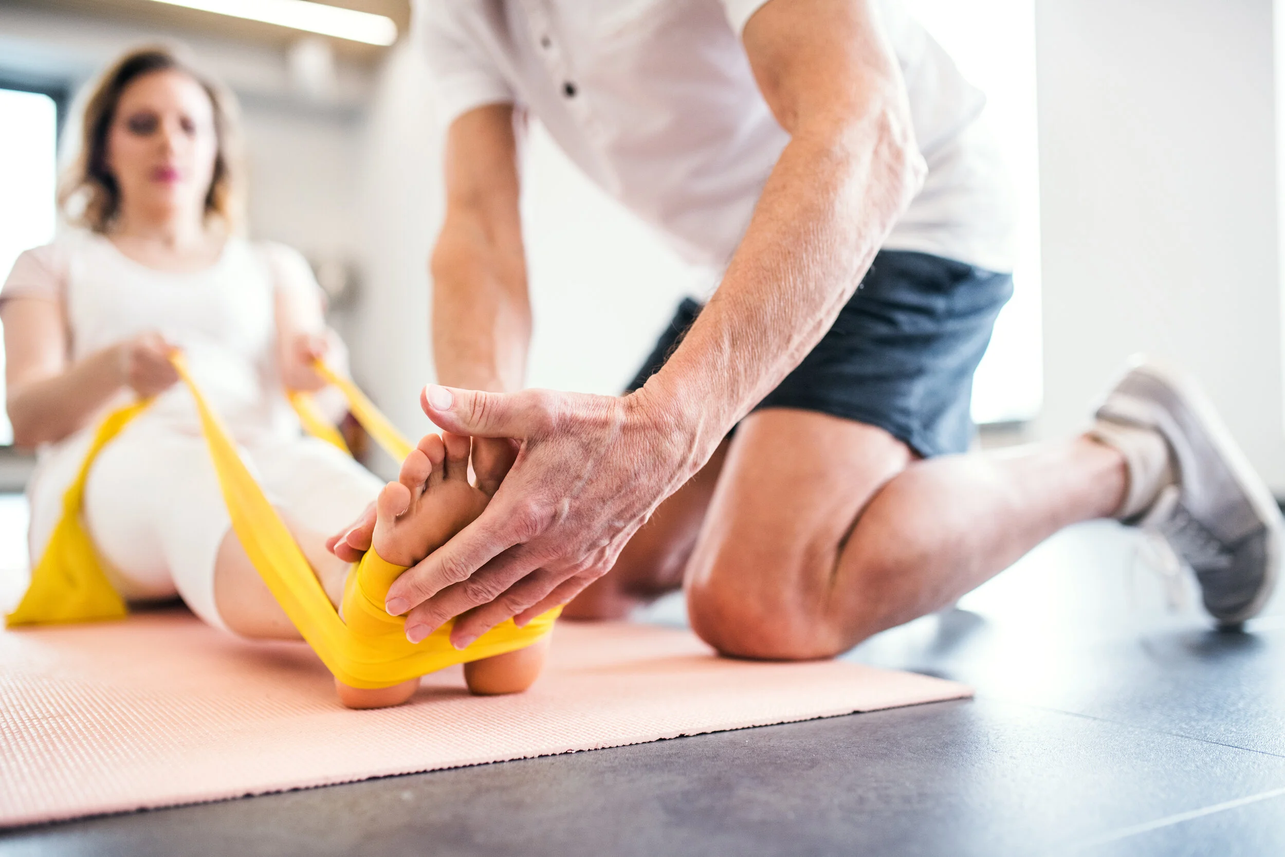
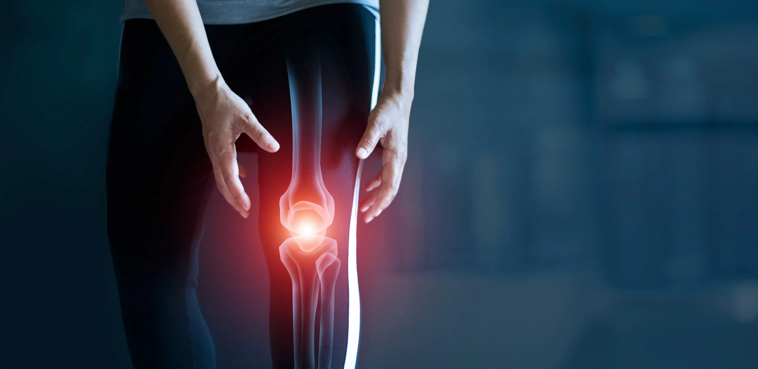
Physical Therapy versus Glucocorticoid Injection for Osteoarthritis of the Knee
Abstract
BACKGROUND
Both physical therapy and intraarticular injections of glucocorticoids have been shown to confer clinical benefit with respect to osteoarthritis of the knee. Whether the short-term and long-term effectiveness for relieving pain and improving physical function differ between these two therapies is uncertain.
METHODS
We conducted a randomized trial to compare physical therapy with glucocorticoid injection in the primary care setting in the U.S. Military Health System. Patients with osteoarthritis in one or both knees were randomly assigned in a 1:1 ratio to receive a glucocorticoid injection or to undergo physical therapy. The primary outcome was the total score on the Western Ontario and McMaster Universities Osteoarthritis Index (WOMAC) at 1 year (scores range from 0 to 240, with higher scores indicating worse pain, function, and stiffness). The secondary outcomes were the time needed to complete the Alternate Step Test, the time needed to complete the Timed Up and Go test, and the score on the Global Rating of Change scale, all assessed at 1 year.
RESULTS
We enrolled 156 patients with a mean age of 56 years; 78 patients were assigned to each group. Baseline characteristics, including severity of pain and level of disability, were similar in the two groups. The mean (±SD) baseline WOMAC scores were 108.8±47.1 in the glucocorticoid injection group and 107.1±42.4 in the physical therapy group. At 1 year, the mean scores were 55.8±53.8 and 37.0±30.7, respectively (mean between-group difference, 18.8 points; 95% confidence interval, 5.0 to 32.6), a finding favoring physical therapy. Changes in secondary outcomes were in the same direction as those of the primary outcome. One patient fainted while receiving a glucocorticoid injection.
CONCLUSIONS
Patients with osteoarthritis of the knee who underwent physical therapy had less pain and functional disability at 1 year than patients who received an intraarticular glucocorticoid injection. (ClinicalTrials.gov number, NCT01427153. opens in new tab.)
Gail D. Deyle, D.Sc.,
Chris S. Allen, D.Sc.,
Stephen C. Allison, Ph.D.,
Norman W. Gill, D.Sc.,
Benjamin R. Hando, D.Sc.,
Evan J. Petersen, D.Sc.,
Douglas I. Dusenberry, M.S.,
and Daniel I. Rhon, D.Sc.
https://www.nejm.org/doi/full/10.1056/NEJMoa1905877
Pelvis Feel Out of Whack?
Lateral Pelvic Tilt: Its Cause and How to Fix it
What causes a lateral pelvic tilt?
The simple answer is that a lateral pelvic tilt is caused by tight muscles on one or both sides of the pelvis. These tight muscles hold the pelvis in a tilted position.
In a laterally tilted position, the pelvis assumes a position where one side appears higher than the other.
Some people will refer to it as a “twisted pelvis” or “rotated pelvis” because along with one side being higher, their pelvis may feel twisted. In fact they are correct, their pelvis is twisted and tilted.
So while the simple answer is technically true, it’s only a partial answer to a more complicated answer.
The more complicated answer, and one that virtually no one realizes, is that a lateral pelvic tilt IS NOT a local pelvis and muscle issue.
A lateral pelvic tilt is a total body issue!
Once you actually understand that lateral pelvic tilt is a total body issue, resolving a lateral tilt becomes easier because you don’t have to search for any one muscle in particular that is causing the problem.
Lateral Pelvic Tilt: My Struggle
The traditional thinking of a lateral pelvic tilt is that there is one or two muscles that can be identified as being tight, and that the tilt is a result of these tight muscles pulling the pelvis in one direction or another.
For a long time I thought this, too.
After all, this is the paradigm through which much of physical therapy operates: stretch tight muscles and strengthen weak ones.
Unfortunately, this understanding of a lateral pelvic tilt, while completely understandable, is seriously flawed and generally leads people on a wild goose chase.
We search in vain for a tight muscle to stretch or massage that will release the tilt and return the pelvis back to its normal resting state. Sometimes we have a little success, and feel some relief, but then the tilt returns.
I went through this for years!
The two pictures above were both taken in 2011 after a particularly vicious lower back spasm that kept me from straightening my back for two entire days. The pain was horrendous.
I could not get up off the floor. Trying to straighten my back would just result in more spasm. So I stayed on the ground for two days.
Once the spasm resolved, I remained with a tilt as seen in the picture on the right. From my notes (I started taking extensive notes of my physical experiences during this time) I got out of the first tilt by stretching my right QL.
Unfortunately, I spasmed again into the position on the left.
It’s like my body was playing ping-pong with my pelvis.
Still in pain, I started researching every possible solution.
I tried physical therapy, which did nothing. Strengthening the core is often nothing more than a cruel joke.
Stretching and massage gave me a bit of relief, but nothing lasting.
After many months of pain and confusion, much of my tilt had gone away because the muscles had finally relaxed, but my left SI joint was still killing me.
Also, oddly, I realized I had one leg shorter than the other and this was tilting my pelvis and stressing out my SI joints. Was I born with a leg length discrepancy?
More confusion!
I put some paper towels in my shoe to make my right leg longer, effectively leveling out my pelvis, and the pain would go away.
But unless I wanted to live with paper towels in my shoe for the rest of my life, I had to address the underlying structural dysfunction.
Two years later, the situation hadn’t changed and I wrote this on my blog:
Two years of physical misery and no one could get to the origin of the problem.
Then I took Myokinematic Restoration from the Postural Restoration Institute and discovered the truth about lateral pelvic tilts.
Lateral pelvic tilt is caused by your pelvis being stuck in an asymmetrical position due to faulty sensory processing.
The typical asymmetrical position consists of a left side of the pelvis rotated forward compared to the right side (in PRI terms, a left AIC pattern).
In this position, some muscles are constantly short and tight and some muscles are constantly stretched and weak.
To see how the wrong eyeglasses will put me into a lateral pelvic tilt and give me a scoliotic looking back, check this page out.
Lateral Pelvic Tilts are Total Body Issues
Let me begin by stating this clearly: you can not stretch or massage your way out of the underlying pelvic asymmetry that is causing the lateral pelvic tilt.
The “tight” muscle that is holding your pelvis in a tilted position (quite often your right or left quadratus lumborum) did not just one day decide to get tight.
Muscles do not tighten independent of the rest of the body.
While it is a local muscle (such as a right QL) that is holding the pelvis in a tilted position, that local muscle was forced into that situation by the asymmetric resting position of the pelvis. The QL is more a victim than perpetrator.
As an example, let’s examine the right QL, the position it can be forced into, and it’s role in a pelvic tilt.
The Right QL
The quadratus lumborum is not actually considered a back muscle, it’s considered a posterior abdominal muscle. But really, that is not important at all.
What is important is the QL’s three attachment sites:
at the top of the pelvis
transverse processes of the lumbar vertebrae L4,L3,L2, and L1.
bottom portion of the last rib.
The QL also has a direct connection and influence on the SI joint because the ilio-lumbar ligament has its embryological origin in the QL, and this ligament plays a vital role in stabilizing the SI joint.
In addition, the ilio-lumbar ligament has a plethora of nociceptors and mechanoreceptors, so it will be quite sensitive to abnormal pelvic mechanics.
When the QL is tight, more often on the right side (in the left AIC pattern, our bodyweight is shifted to the right), it can contribute to abnormal pelvic mechanics (and thus intense pain) in the form of a lateral tilt as it pulls the pelvis and ribcage closer together on that right side. Hence it’s nickname, the “hip hiker”.
But why on earth is that right QL tight?
Out of all the muscles in the body, why does this muscle seem to get tight so often? Doesn’t it seem odd that this lonely right QL just gets tight out of nowhere?
It should seem odd, because that isn’t what happens.
Pelvic Orientation to the RIght
The right QL only gets tight because it is forced into that position by the two structures it attaches to, the pelvis and the ribcage (remember it attaches to the 12th rib, so action of the ribcage influences the QL, as well.)
The QL’s job is to side-bend, and then stabilize, the pelvis, ribcage, and spine to the same side.
So a right QL will bring the right pelvis and right ribcage closer together by sidebending the spine to the right.
All this sidebending and stabilization produces a pelvis that is oriented to the right.
If the right QL is tight, it means the entire pelvis is oriented to the right.
A right QL will not be tight if the pelvis is oriented to the left. This is important to understand, otherwise the remedy won’t make sense.
Muscle Behavior
Muscles have attitudes and behaviors just like our personalities do.
Once a muscle’s behavior has become habitual, it doesn’t give up its habit easily. In this case the right QL is thinking “I’m doing exactly what you asked me to do, keep the right hip and ribcage stable”.
If you never shift your weight fully to the left, or something is preventing you from shifting your bodyweight fully to the left, why would the right QL ever turn off?
It will only fully turn off when you fully shift your weight to the left because if your weight is on the left foot, the right QL has no reason to stay on. Your left QL will activate when you shift your weight to the left.
The right QL will turn off at that time.
The pictures below show what a lateral pelvic tilt caused by a tight right QL would look like. When my weight is on my right leg, my body looks ok because in reality my right should be slightly higher when my weight is on the right foot.
When my weight is on the left leg, but my pelvis remains oriented to the right, my upper body has to awkwardly compensate.
In addition, am I truly on my left leg?
This is a huge leap in understanding right here. You can put your weight on your left leg without achieving a true left leg weight-bearing position.
But this is “fake” left leg weight-bearing.
It’s phony.
It’s fugazi.
True left leg weight-bearing requires true left leg and hip musculature to activate appropriately. If these muscles fire appropriately then your pelvis will orient TO THE LEFT when your weight is on your left foot. It will happen automatically.
In other words, your pelvis WILL NOT remain oriented to the right if you are truly on your left leg, and your right QL will not remain tight if you are truly on your left leg because the right QL turns off when your weight is truly on your left leg.
Weight on right foot, right pelvis orientation. Normal mechanics.
Weight on left foot, pelvis orientation still to the right = fake, phony, fugazi left weight-bearing position
This is what the pelvis should look like in right and left weight-bearing.
But this is not happening when you are stuck in a pelvic tilt!
Weight on the right foot, right pelvis and ribs closer together, pelvis oriented right.
Weight on the left foot, left pelvis and ribs closer together, pelvis oriented to the left. This should be normal.
Because the position of your pelvis and ribcage DO NOT CHANGE when you shift your weight to the left (because of the left AIC pattern), the right QL is habitually stuck in a shortened and tightened position.
It’s doing exactly what it is supposed to do!
If your weight is on the right foot, your right QL should be stabilizing your right side by bringing the right pelvis and right ribcage closer together. That’s normal right stance mechanics.
The right QL will continue to stay “on” if your pelvis stays oriented to the right even when your weight is on your left foot. That is not normal left stance mechanics. It’s fake left stance.
To fix the situation you have to attain a new pelvic behavior that orients the pelvis to the left when your weight is on the left foot.
It’s as simple as that.
The Solution
The only way to eliminate the underlying structural issue that is leading to lateral pelvic tilt is to reposition the pelvis into a more symmetrical resting position,
Once pelvic repositioning has occurred, you then train your body to establish and stabilize itself in left stance.
For people who already understand PRI, this means that you need to strengthen the left hamstring, left IC adductor, left glute medius, and left internal obliques using PRI exercises. All these muscles stabilize our body in left stance.
For starters, You can try the exercises on this page.
I also offer Skype consultations if more help is needed. You can e-mail me Nealhallinan@gmail.com for more information.
Or just start with the one below.
This single leg exercise is designed to re-orient your pelvis and ribcage to a *neutral position* so that they are no longer oriented to the right. The left hamstring is what pulls the pelvis into that neutral position and the left internal obliques (via full exhalation) bring the ribs over to the left.
Using a small ball between your knees can help feel the proper muscles.
You should feel your left hamstring working.
Don’t “push into the wall” with your left foot. You need to keep the left foot flat and “pull down” with your heel towards the ground (although the foot should not move).
Make sure you exhale completely so that you feel your left ribs coming “down, back, and in”.
Some people have both sides of their pelvis rotated forward (PEC pattern) and/or may feel like the right side of their pelvis is rotated forward compared to their left.
While this is probably not what is actually happening, you can try a two legged version of this exercise by keeping both feet on the wall and not including any hip shift. In this case you would be recruiting both hamstrings.
If you have any questions, just leave them in the comments section and I’ll answer as soon as I see it.
Better PT Access Leads to Better Outcomes
Better PT Access Leads to Better Outcomes
POSTED ON: November 14, 2019TOPICS: chronic pain, health insurance, insurance, pain, physical therapy
Because of School of Public Health research, a major insurer has changed its benefits to make it easier for patients to access physical therapy and chiropractic care for low back pain.
Research published in the journal Physical Therapy is the newest of three studies by the team, finding that unrestricted direct access to physical therapy leads to less health care utilization and lower costs. The other two studies found that insurance policies affect the likelihood that someone with low back pain will go straight to a physical therapist or chiropractor for treatment rather than a primary care physician (PCP), and that going straight to a physical therapist or chiropractor reduces opioid prescriptions.
The research has led UnitedHealthcare to introduce a new benefit where patients do not have to pay for their first three visits for physical therapy or chiropractic care to treat low back pain.
“This is a wonderful outcome of our work, which will likely shape the way consumers of care make their own choices regarding which provider to see,” says Lewis Kazis, professor of health law, policy & management, who served as senior author of the most recent study, and senior and lead author, respectively, of the other two studies.
Kazis and colleagues conducted the research with OptumLabs, which provided claims data on a study cohort of nearly 60,000 patients who had new-onset low back pain between 2008 and 2013 (OptumLabs is part of UnitedHealth Group.).
For the newest study, the researchers compared health care utilization among individuals who first saw a physical therapist for low back pain in states with unrestricted access to physical therapy and in states with provisional access. The researchers found that the individuals in states with only provisional access were billed for more health care utilization within 30 days, such as getting X-rays and visiting physicians more frequently.
The researchers then compared low back pain–related costs among patients who first saw a physical therapist and those who first saw a PCP for the condition. In provisional-access states, patients who went to PT first had 25-percent higher costs at 30 days and 32-percent higher costs at 90 days than those who saw a PCP first. But in states with unrestricted access to PT, patients who first went to PT had 13-percent lower costs at 30 days and 32-percent lower costs at 90 days than those who first saw a PCP.
All together, Kazis says, the three studies show that easier access to PT and chiropractic care for low back pain leads to patients seeking such care first, rather than going to a PCP first, and that doing so reduces health care utilization, costs, and opioid prescriptions.
It Takes All of Us
Collaboration Between Physicians & Physical Therapists Is the Future of Medicine
It's a well-known fact that as elderly people age, physical function declines. The natural deterioration of musculoskeletal systems in older adults can dramatically reduce quality of life, but perhaps even more importantly, it is strongly associated with increased hospitalization, disability, dependence, and mortality rates.r' This puts a significant strain on both elderly people and the health care systems serving them across the world, especially as the number of elderly adults increases.s With this in mind, it's vital that the American medical establishment recognize the links between functional limitation, disability, and illness in the elderly and develop methods to help patients maintain physical function as they get older. Of course, with a wide range of health conditions that contribute to physical decline, including sarcopenia, obesity, chronic pain, and cognitive decline, it's impossible to recommend a one-size-fits-all approach. However, as the medical establishment seeks to develop and refine a multilateral treatment strategy, preventative physical and occupational therapies have emerged as powerful tools to simultaneously mitigate a wide range of risk factors for functional decline in the elderly.'
Where Physical Therapy Fits In
Physical activity is strongly linked to overall health, longevity, and well-being in individuals ofall ages, but research indicates that it may become even more important as patients move past the age of 65.'But how do we encourage elderly adults to stay physically active as their muscles deteriorate, chronic disease enters their lives, balance becomes an issue, and cognitive decline keeps them sedentary? By implementing preventative physical therapy intervention in elderly patients' regular treatment plans, we can address each of these problems at the root and help patients maintain the baseline health levels required to stay active. In one 2015 study, 41o community-dwelling adults over 75 years of age were split into three groups. Two of the groups went through a functional task exercise or preventative physical therapy program, both administered by physical therapists, while members of the control group continued to receive their usual levels of care. Researchers asked participants to self-report their functional abilities on two questionnaires, before and after the year-long trial. While both the experimental and control groups declined in physical function over a year, the experimental group's rate of functional decline was slowed by two-thirds.' This research supports a growing body of evidence indicating the efficacy of physical therapy intervention in slowing down functional decline in the elderly. One 2016 review concluded that "supervised resistance and/or aerobic PA interventions significantly [improve] performance-based, composite PF outcomes among community dwelling older adults" and that supervised exercise programs administered by professionals can improve long-term physical function.'The more minutes of exercise participants engaged in per week, and the longer they adhered to the exercise program, the better the patients' outcomes were.
Proactive Intervention Is Key
We're guessing that the evidence described above comes as no surprise to most physicians. Physical therapy encourages physical activity by design, and physical activity has long been linked to health and well-being. But it's important for medical practitioners to note that exercise intervention is more effective before an elderly patient is injured than after. Mortality rates skyrocket after a patient falls the first time, and subsequent falls become far more likely. Therefore, it's recommended that all at-risk elderly patients be screened for functional ability and prescribed preventative physical therapy as needed. Mobility performance analyses, such as screens examining walking speed and balance, are excellent tools to find the patients who can benefit most from preventative therapy.'By steering patients who exhibit significant functional decline toward a comprehensive physical therapy program, we can dramatically reduce the individual and collective stress that aging poses to our population.
Chase |D, Phillips L|, Brown M. Physical Activity Intervention Efects on Physical Function Among Community-Dwelling Older Adultsr A Systematic Review and Meta-Analysis. Journal ofAging and Physical Activity. 20 I 7;25( l): r49'170. doi:10. t r23ljapa.20r6'0040. '1Siemonsma PC, Blom JW Hofstetter H, et al. The efectiveness offunctional task exercise and physical therapy as prevention of functional decline in conlmunity dwelling older people with complex health problems. BMC Geriatrics. 2018;18(l). doi:10.1186/s12877-018-0859-3. Reeves D, Pye S, Ashcroft DM, et al. The challenge ofageing populations and patient frailty: can primary care adapt? BMl. 2018i362:k3349. doi:10.1 136/bmj.k3349. aAnton SD, Woods AJ, Ashizawa T, et al. Successful aging: Advancing the science ofphysical independence in older adults. Ag€ing Research Reviews. 2015;24:304-327. doi:10.1016/j.arr2015.09.005.
KEEP MOVING: Is Exercise the Answer to Lower Back Pain?
When patients report low back pain, it has often already impacted their quality of life. They are challenged by daily tasks and common activities. When pain begins to manifest, a person's first response may be to take over-the-counter pain medication and rest. This, however, may only increase the duration or intensity of the pain.
Thus, it becomes the challenge of the clinician to communicate to patients that over-the-counter pain medication and rest may not be the answer to lower back pain - or back pain in general. With proper diagnosis (ruling out spinal issues, such as herniated discs), the best instruction for the patient may be increased movement. This could include a daily exercise program.2'3
It becomes crucial to educate and evaluate patients on proper exercise and stretch techniques. Clinicians should evaluate movement and ensure the patient is exercising properly and safely and getting the benefits of the exercises, the primary one being a reduction in low back pain (LBP).
Numerous studies have confirmed that movement, including high-quality aerobic exercises, calisthenics, hydrotherapy, and even daily walks can be effective at reducing pain and restoring function. This includes both clinic-based programs and at-home exercises. Individually designed strengthening and stabilizing regimens have been shown to be just as or more effective than other conservative treatments.3,4,5
There has been some debate as to whether or not exercise is just as effective as simply staying active (e.g. the patient resuming their normal daily activities or prior exercise). For chronic LBP sufferers, a structured exercise program may be more effective.
Patients with acute LBP typically respond better when resuming their normal daily activities and not just "taking it easy" or lapsing into a sedentary lifestyle in an effort to heal or avoid the pain. An official diagnosis should determine the course of action regardless. 5
Many people who experience chronic low back pain also experience issues related to trunk strength, diminished flexibility, and limited endurance. Developing an exercise program that targets these areas may be advisable. By improving strength, flexibility, and endurance, patients should begin to see a reduction in pain.
In studies of chronic LBP, subjects who participated in an exercise intervention, namely strength/resistance and coordination/stabilization interventions, were noted for having the greatest impact on reducing low back pain. Clinician monitoring rs recommended.
LBP is a group of conditions that affects millions of Americans. With improved education and intervention, we can better address LBP with movement-based programs. One major hurdle to overcome is patient fear - fear of pain, which can lead to a short-term or long term sedentary lifestyle. Movement is the answer to many forms of LBP. If you have a patient who is suffering from a low quality of life due to LBP, contact our office today to see if physical therapy is the right choice.
1. Costa L da CM, Maher CG, McAuley lH, Hancock Ml, Smeets, RJEM. Self-emcacy is more important than fear of movement in mediating the relationship between pain and disability in chronic low back pain. European fournal of PaiIr. 201 I;15(2);2I3-219. doi:10.1016/j. ejpain.20l0.06.0l4
2.Cote JN, Hoeger Bement MK. Update on the Relation Between Pain and Movement: Consequences for Clinical Practice. The Clinical lournal ofPain 2O10;26(9):754-762. doi: 10.1097/ajp.0b013e3l8le0l74f
3. Hayden fA, van Tulder MW, Malmivaara AV Koes BW Meta-Analysis: Exercise Therapy for Nonspecific Low Back Pain. Annals of Internal Me dicine. 2005i142(9\ 765-775. doir0.732610003-4819-142-9-200505030-000 I3
4. Liddle SD, Bdter DG, Gracey JH. Exercise and chronic low back pain: what works? Pain. (2004);107(l):w176-190 doi:10.1016/j.pain.2003.10.017 5
5.Searle A, Spink M, Ho A, Chuter V Exercise inteilentions for the treatment ofchronic low back pain: a systematic rcviewandmeta-analysisofrandomisedcontrolledtrials.ClinicalRehabilitation 20l5;29(12\:1155-1167, d,ol lO.l 17 7 / 026921. 5 5 I 557 O37 9
MRI PRIOR TO CONSERVATIVE THERAPY OFTEN UNNECESSARY FOR ATRAUMATIC SHOULDER PAIN
Originally published on Journal of Shoulder and Elbow Surgery
KEY FINDINGS
This prospective study examined the value of MRI in the initial management of patients with atraumatic shoulder pain, suspected cuff tendinopathy and minimal to no strength deficits
After receiving at least two months of conservative therapy, only five of 51 patients (90%) underwent surgery over an average follow-up period of 28 months
The surgery cohort underwent an operation at an average 68 days after MRI, indicating that they did not need MRI at initial presentation
In this population, surgeons should consider acquiring MRI only after conservative therapy fails
Rotator cuff tears (RCTs) are quite common in otherwise healthy populations (34% in a study that included partial tears published by University of Miami researchers in The Journal of Bone and Joint Surgery), and the prevalence increases with age. But despite this fact, there are no clear guidelines about their evaluation and management.
Based on clinical examination alone, it's exceedingly difficult to identify the type of shoulder injury in a patient who has pain but only slight physical deficits. U.S. clinicians often order an MRI at the initial presentation of suspected shoulder tendinopathy or shortly thereafter.
Based on data from a prospective study, Scott D. Martin, MD, director of the Joint Preservation Service within the Department of Orthopaedics Sports Medicine Center at Massachusetts General Hospital, and colleagues now call into question the use of MRI before a trial of conservative management in patients with atraumatic shoulder pain, minimal to no strength deficits and suspected cuff tendinopathy other than full-thickness tears. Their report appears in the Journal of Shoulder and Elbow Surgery.
Study Details
The researchers followed 51 adults who had a chief complaint of atraumatic anterolateral shoulder pain, strength test minimum score of 4 or 5, and screening radiographs exhibiting no to mild glenohumeral arthritis and no cuff arthropathy.
All patients were suspected to have cuff tendinopathy, but a full-thickness tear was considered unlikely in patients with relatively well-preserved strength testing. They underwent MRI or magnetic resonance arthrography (MRA) at an average of 10 days after presentation.
The patients tried conservative therapy for at least two months including patient education, activity modification, nonsteroidal anti-inflammatory drugs and physical therapy. Those with severe pain were offered an intra-articular steroid injection. If symptoms progressed or failed to improve, patients were given the option of rotator cuff repair surgery.
Need for Surgery
The average length of follow-up from MRI/MRA to chart review was 28 months. Over this period only five patients (10%) went on to surgical intervention. Thus, 46 patients had premature MRI/MRA that did not affect management.
Risk Factors for Surgery
Four of the five patients requiring surgery had full-thickness tears on MRI, and conversion to surgery was significantly associated with such a finding (P ≤ .001). There was borderline correlation between surgery and age (r = 0.274; P = .0518).
Thirty-three patients had concomitant labral pathology, but it was not a statistically significant risk factor for surgery.
A More Reasonable Approach
The patients who had surgery received it an average of 68 days after MRI/MRA. In retrospect, they could have tried 60 days of conservative therapy, undergone an MRI if symptoms persisted or progressed and received surgery within the same time frame. This seems to be a more reasonable approach, as it would have yielded similar results and posed much less financial burden to patients and the health care system.
Value in health care has been defined as outcome relative to costs. MRI is an exceptionally powerful diagnostic tool, but to translate into value, it must provide actionable information that influences management.
The Whole Body Fix
By: Katie McDonald Neitz (http://m.runnersworld.com/person/katie-mcdonald-neitz)
Chronically injured and disheartened, a Runner's World editor sought holistic help from a team of therapists. Her diagnosis (sleeping glutes?) and hard-won lessons (master the clamshell!) can help you, too, stay healthy, happy, and on the road.
Image by: Reed Young Published: February 27, 2014
I'm lying facedown on an exam table at a state-of-the-art running clinic in New York City, about to perform a basic exercise for professional analysis. "Okay, Katie, I'd like you to lift your right leg in the air, using your glutes," says Colleen Brough, P.T., M.S., the physical therapist who's there to check my strength and form. No problem, I think. She places her hand on my right hamstring–my achy, troublesome one–as I lift and then lower my leg back down to the table with minimal effort and an attitude of That's all? "You contracted your hamstring as well as your back," Brough says gently. "Try again, but this time, power the move with your glutes by squeezing your butt before and while doing the lift." Okay, got it. Simple enough. But it isn't. Impossible, actually. I lie there motionless, slowly coming to the realization that clenching your face doesn't help you clench your butt cheeks. Forget lifting the leg. I am entirely unable to activate my glutes, a fact Brough describes with one cruel but apt word: "Astonishing."
What might be even more astonishing is that I'm Runner's World's Mind+Body editor. When a person's job requires talking with top sports-medicine docs and physical therapists to provide injury prevention and treatment advice for runners, you might assume said person would be a reflection of her work: a happy, muscle balanced, injury-free runner. Instead, I've been struggling with the same strained hamstring for six years. I've tried every technique recommended in these pages: massages, Active Release Technique, Graston Technique, and chiropractic treatment. And everything has made me feel better–temporarily. My improvements are always short-lived.
THE MISTAKE: RUNNING AND RACING WITH A SERIOUS WEAKNESS
While my race times aren't very impressive, what is impressive is that I have been able to run half-marathons at all, with no help from my glutes. My glutes– specifically my gluteus medius and gluteus maximus muscles–are asleep, even on the run, and perfectly fine with having my hamstrings, hips, back, and abdominal muscles step in to carry the load. And that's a problem. A common one, it turns out. Each of the 11 physical therapists and exercise physiologists I spoke with for this piece named inactive glutes as the top weakness they see in runners (with weak lower abdominals as a close second).
Why? One theory is that we park our butts too much. "Many people drive to work, then sit all day, then drive home and sit more," says physical therapist Craig Souders, P.T., the 2:52-marathoner who is overseeing my rehab at Lehigh Valley Health Network in Allentown, Pennsylvania. "That's not the function of the core, which includes your glutes. It's designed to move. So we see something that's like disuse atrophy. It's this pattern where the muscle is turned off. And when it's turned off, it's hard to get it back on."
THE FIX: GET HELP
You took extra rest days, you foam-roll regularly, you dialed down your mileage, and still you hurt? A chronic soreness or achiness that doesn't go away is a sign of an injury (or an emerging one) that deserves professional attention. Admitting that you need help is step one. Step two is getting the right kind of help. You could go to your regular doctor, chiropractor, or massage therapist and get lucky–he or she may have experience treating runners and understand our unique biomechanics and the common muscle weaknesses and compensations that often occur in our bodies. Or your professional might treat the symptom of your problem (in my case: a sore hamstring) without discovering the underlying cause of your injury (in my case: inactive glutes). Focusing on the symptom, and not the true source of the problem, isn't an effective solution. This is the value of visiting a running clinic, like RunSmart, that takes a holistic approach to injury prevention and diagnosis.
THE MISTAKE: ACTING LIKE A STUD
I pride myself on being a well-rounded athlete. I run, swim, and spin a few times a week. And when I strength-trained, I had the attitude that I was too "advanced" for simple exercises, like clamshells. Instead, I did complex movements like burpees with box jumps. I figured all this cross-training had made my body strong and balanced. And so I was excited to do the Functional Movement Screen (FMS) at the RunSmart clinic, which measures head-to-toe strength and flexibility and is designed to detect muscle imbalances via a series of seven exercise tests. This would be my opportunity to shine–so I thought. A person is considered vulnerable to injury if he or she earns fewer than 14 points out of a possible 21 on the FMS. My score: 12.
I imagine I'm not the only runner with a false sense of confidence about my fitness. It's easy to assume that because you are capable of running 13.1 miles in one shot or even 13.1 miles in one week, you are strong. And you are, for sure. But that's not all that matters. "You can get strong doing something incorrectly– using a dysfunctional movement pattern," says Alison Peters, M.S., a clinical exercise physiologist at NYU Langone who conducted my FMS test. "Your body is designed that way–to enable you to do the movement, even if it's not being done correctly. The problem is that you'll eventually get injured from performing the move that way."
THE FIX: FOCUS ON THE BASICS
To address specific weaknesses and correct flawed patterns, you've got to take a step or two back and home in on the exact area that needs improvement. If your glutes aren't firing, for instance, you can do squats all day long and you'll just continue to work (and likely strain) whatever muscle group covers for your comatose rear. Instead, you need to isolate your true problem spot. This may require checking your ego at the gym door and doing exercises you might have once deemed too remedial for you. "It's tough to convince runners that basic strength exercises, like clamshells, are important," Koo says. "But if you jump into complex moves without the necessary strength or range of motion, you're almost setting yourself up to fail. Maybe not immediately, but eventually, it'll lead to injury. You need to work on the basics and focus on quality over quantity."
This can be challenging for ambitious, numbers-driven runners, who'd rather do 20 leg lifts than 10–but if you are doing them with poor form and relying on the wrong muscles in order to crank out a high number of reps, you aren't doing yourself any favors. "Practice doesn't make perfect; perfect practice makes perfect," Koo says. "If you do each rep as perfect as possible, you'll notice the muscle burns, it's challenging, and you'll feel like it's worth your time." Indeed, says Brough. "You might be able to do 100 clamshells and think it's an easy exercise," she says. "But can you do it while maintaining a lower abdominal contraction and while activating your glutes? When you recruit the right areas, suddenly an 'easy' exercise isn't so easy."
THE MISTAKE: TAKING A HALFHEARTED APPROACH TO RECOVERY
During all my attempts to cure my hamstring, I've kept training for races. My top priority would be my running–not my rehab. I remember one physical therapist very gingerly suggesting that I'd likely see better results if I didn't run 10-milers every weekend. I stuck my fingers in my ears and started singing, "La, la, la, la! I can't hear you!" Okay, I didn't actually do that, but I might as well have.
It's also probably no coincidence that during the six years I've been battling my hamstring, I had two kids. That means my sleep quantity has been limited, and the quality of sleep I have gotten has been poor. "There is good research that indicates that anything less than seven and a half hours a night predisposes you to injury," says Laura Dunne, M.D., who is a primary-care sports-medicine physician at OAA Orthopedic Specialists in the Lehigh Valley, Pennsylvania, and also my doctor. Yet, regardless of how little sleep I might have gotten, I'd still get up with my alarm (predawn, even on weekends). The idea of moving a run to a time when I might be better rested made me cranky. I also made the mistake of seeking a cure from massage therapists, chiropractors, and other practitioners without taking enough personal responsibility for my own recovery (see "Trial and Error," page 53). I suppose I thought that I could simply lie on a table and have a professional render me injury-free. My lazy mind-set had to change. Souders has helped me flip this mental switch. "If you want to get better, you need to attack your rehab," Souders told me at my first appointment. In other words, put just as much focus and energy into it as you would a training run and accept it as a serious mission. I'm learning, for example, that it's better to do a three-mile run than a five-miler, and spend those extra 20 minutes on the exercise mat.
THE FIX: PRIORITIZE YOUR REHAB
Just as nailing a PR isn't easy, recovering from an injury isn't easy–it takes dedication and hard work. And just like with your training, you can't expect to get better if you give it less than your best. "I tell my patients to channel the energy and frustration that you can't run or can't run as much as you want into your rehab," Souders says. "Get mad! Do 5,000 clamshells a day because you're so mad. If you commit to that and do that, then when you get back to running, you'll see real progress."
It's also important to be mentally engaged in your recovery. "It's very easy to go into mindless mode," Souders says. "Let me play with the kids, watch TV while I do my exercises, and the next thing you know, you're really not feeling it. You're not activating the right muscles, and you're not feeling the burn." To properly train your muscles and prepare them for the rigors of running, you've got to think of rehab as endurance training, and work your muscles until they are truly tired–not until you've simply tired of doing the work.
And as hard as it is, in your busy, time-crunched life, sleep shouldn't be the first thing to go. "We live in a running culture where if we have a lot going on at work and with the kids, we'll still take on marathon training and just sleep less," says Souders, himself a father of two who is training for the Boston Marathon in April. "But the body repairs itself when you sleep, so if you cut that out, there can be effects." Figure out a balance–whether it's going to bed an hour early when you have an early run planned, or dialing down your training and focusing on, say, a 10-K race goal instead of a half-marathon, when you know your time–and sleep–is limited.
THE MISTAKE: LOOKING FOR A QUICK FIX
Six weeks after I started physical therapy, I returned to the RunSmart clinic for a follow-up evaluation. In that period of time, I saw my therapist only three times. But I was religious about my homework: I did many, many reps of clamshells, leg lifts, and bridges daily–sometimes twice a day. Still, neither the staff nor I expected a dramatic transformation after such a short time, but any sign of improvement would indicate that my therapy was on the right track, reward me for my efforts, and motivate me to keep at it. I thought my FMS score, which measures strength and flexibility, might move up a point or two. Instead, it jumped from 12 to 17. The results of my treadmill analysis were just as striking. My pelvic drop, which was significant at initial testing, was now within a healthy range. Most impressive, perhaps, was that my hamstring didn't hurt. At all. And my therapy hasn't involved any hamstring stretching or massage–just lots of glute work. The old me would have been so pleased with this feedback I'd likely declare myself "cured." The new me, however, realizes I've made good progress, but I'm still very much a work in progress.
All the clamshells in the world aren't going to produce any long-lasting carryover to my running until I take the new muscle recruitments I've been practicing in rehab and put them into effect when I run. This is a critical step, but one that hasn't always been taken. "We found that runners would finish their rehab and be strong, so we'd send them out on the road," Brough says. "But they'd go back out and their pain would still be there. We now know that it's not until you incorporate real-time training cues into your runs–you teach your muscles to be recruited while you are running–that you see a difference." To do that, RunSmart prescribed me two running drills I'll incorporate into my routine once my physical therapist says I'm ready. One focuses on glute recruitment: As I run, I'll focus on squeezing my butt each time I push off with my foot; the second focuses on developing a midfoot strike with a forward lean, which will help me run more efficiently (click here for more detail). To stay healthy, it's recommended that I continue to do these drills regularly–even when I am truly healed.
THE FIX: BE COMMITTED
When you've been running with flawed biomechanics for years, you can't expect an overnight fix. And even when you are declared healed, your faulty habits are something you need to be cognizant of–if you want to avoid another breakdown. "You are going to have these patterns that you tend to fall into," Brough says. "So if you tend to have excessive pelvic drop, that will tend to happen on your last mile or on that tough hill. Those are times to call upon these running cues–contract your lower abs, push off through your glutes–to help you prevent those issues." Souders agrees. "Let's say you run 40 miles a week. What if you make that 36 miles a week, and in that extra time you have, you do drills and exercises that will work your weak spots? The work isn't over when you return to sport. That's when you need to focus on stopping this from happening again."
Living With Arthritis?
21 Tips from Physical Therapists to Make Arthritis More Manageable
Physical therapy can be an important part of treatment for many people with arthritis. Here’s the advice PTs wish they could give everyone.
By the time you get diagnosed with arthritis you may have a panel of different doctors and nurses who’ve helped you along the way, but there are other medical professionals who can have a big impact on your health and comfort — including occupational therapists, mental health experts, and physical therapists.
Physical therapists are best known for guiding people through exercises to help with rehabilitation and manage pain, but they are so much more than a personal trainer, says Lauren Lobert, a physical therapist and owner of APEX Physical Therapy. A good PT will employ a variety of techniques, including targeted exercises and stretches, manual therapy, and biomechanical analysis among other things. They can help you maintain your health long-term by creating a program that you can use out of their office, giving you the tools to manage arthritis flare-ups and improve your daily quality of life, she says.
We talked to a group of physical therapists for their best advice for people managing arthritis:
The Mental Mindset
1. Don’t forget the reason you’re doing this
Physical therapy can at times be painful, frustrating, or even boring — and that’s understandable as you’re pushing yourself more, says Lia Vann, a physical therapist at Midwest Orthopedics at Rush Chicago. But in those moments it’s important to keep your eye on the prize and remember why you’re doing this in the first place. “The goal of physical therapy is to progressively get stronger in order to be able to not just move more, but to move better,” she explains. A good PT will keep an updated list of your goals and check in with you about your progress.
2. Surgery is not a cure-all
Joint replacement surgery is a necessity for many patients with arthritis. But too many people think it will solve all their mobility problems and their pain will instantly go away, Vann says. Unfortunately the truth is that surgery is not a quick fix. In the initial weeks afterward, things often feels worse before they feel better. One of the best way to help yourself after surgery? Keep your PT appointments, Vann says. “The most positive outcome [from surgery] requires a period of dedicated rehabilitation and we are trained to help you with that,” she says.
3. Ditch the victim mentality
Instead of thinking of yourself as being at the mercy of your arthritis, it’s important to think of your condition as something that can be managed, by you, says Lyndsay Hirst, a physical therapist at Your Pilates Physio. “I tell my clients to imagine they have a tool box and in it are all the things that help their arthritis — things like medication, pain relief, exercise, heat, rest, physical therapy, and so on,” she explains. “Then you feel in control of your illness and can draw out what you need, when you need it.” Need help? A good PT can give you lots of good “tools” for your toolbox, she says.
4. Learn how your joints work
Arthritis patients are often more committed to doing their prescribed physical therapy exercises when they understand how their joints work, says Lauren Peterson, a physical therapist and owner of FYZICALTherapy & Balance Centers. “When your joints move they produce a liquid called synovial fluid, which reduces friction in your joints,” she explains. “This means that your joints might ache a little initially when you start moving the joint, but discomfort should improve with continued movement as you get more fluid to lubricate it.” Your doctor or PT can show you a model or diagram of what your joints look like when they move and how arthritis can change that.
5. You are not your X-rays
Scary-looking scans mean serious pain and disability, right? Not always, Vann says. “I always tell my patients that just because they have osteoarthritis, that does not necessarily mean they are going to have pain,” she explains. “A lot of people get really worried when they are told they have ‘bone on bone’ or have degeneration but just because that is how the joint looks, it does not mean they will have a lot of pain or issues with daily life.” A physical therapist can help you identify any weak spots and give you tips to prevent future pain, she adds. It’s important to talk to your doctor about what your imaging means for your prognosis and whether they indicate any changes in treatment may be needed.
6. Start making changes now
“One of the things I most wish my patients with arthritis understood is that time is of the essence. Arthritis is a progressive and chronic disease,” says Alice Holland, DPT, director of Stride Strong Physical Therapy. Avoiding action or “powering through” can cause further damage to occur. The sooner you start taking steps to deal with and treat your arthritis, the better your prognosis can be.
The Exercise Prescription
7. Motion is lotion
When it comes to soothing painful joints sometimes the best treatment may be the last one you want to do — move it. “Movement is integral in keeping your joints as mobile and limber as possible; stop moving and you’ll see an immediate increase in pain, stiffness, and disability,” says Brittany Ferri, MS, a physical and occupational therapist. “I advise all my clients to keep moving as you normally would, as much as possible, and continue to exercise even through small bouts of pain.” Major pain is a reason to stop, but it’s good to push through a little discomfort, she adds.
8. Learn to love the elliptical machine
When it comes to arthritis, not all exercises are created equal, says Kavita Sharma, of Manhattan Pain & Sports Associates, who specializes in patients with chronic pain. This is especially true if you have osteoarthritis; certain types of exercises can increase the wear and tear on your already damaged joints. “I usually recommend low-impact exercise such as the elliptical machine or swimming, as they are more gentle on your joints,” Sharma explains. “These types of movements are less likely to cause further injury and will maintain or even improve your mobility.”
9. Get pumped
While cardio will keep your joints lubricated and flexible, when it comes to the long-term health of your joints, it’s important to strengthen the muscles that support them, Sharma says. “As you strengthen the muscles that support the joint, you offload the pressure,” she explains. However, it’s important to work with your physical therapist to figure out which exercises are best for your particular type of arthritis. For instance, if your knees are the problem then you’ll want to focus on strengthening your quads, hamstrings, hips, and low back.
10. Strength train without weights
Some people aren’t up to ‘pumping iron’ in the gym and that’s totally fine — there are lots of strengthening exercises you can do just with your body, Hirst says. Pilates is a gentle way to strengthen the muscles that stabilize arthritic joints while improving range of motion and flexibility, she explains. Other options are yoga, tai chi, or bodyweight strength workouts you find online. Just be sure to run it by your PT first so they can make sure they are appropriate for your arthritis, she adds.
11. Do dexterity exercises
Don’t forget your small muscles in your exercises, Holland says. Movement is the best thing you can do for your joints but people often forget that applies to the small joints, like fingers and toes, as well, she says. “If you have arthritis in your hands, do hand dexterity exercises— like opening and closing your hand and picking up small objects — to warm up in the morning,” he says. A PT can help you figure out which dexterity exercises are best for you.
12. Switch up your workouts up
It’s easy to get in a rut with exercise but it’s better for your joints if you can be flexible (ha!) with your workouts, Vann says. “It’s really about finding the right type of exercise and modifying activities so you can keep moving without serious pain,” she explains. That might mean walking one week, swimming the next, and lifting weights in between. Your physical therapist can recommend specific activities that will work best for you at your current pain level, she adds.
13. Don’t overdo it
“Many people with arthritis do too little activity, which can cause their joints to become stiff and painful, but others may be doing too much activity, which can add stress to the joints,” Vann says. (And both things can be true for the same person in different situations!) The whole point of exercising is to help you, so if your activities are so intense that they require hours of recovery afterward or cause major pain flare-ups then it’s time to talk to your PT about making adjustments and cutting back, she says.
14. Get your posture evaluated
Good posture is important for everyone but it’s essential for people with arthritis because poor biomechanics when you sit, walk, or lift can put extra strain and wear and tear on joints, Holland says. “A physical therapist can help to correct your biomechanics,” she explains. “We can help correct your bad habits and postural problems so you wear down your joints less, thereby helping to slow the progression of arthritis and reduce pain.”
The Lifestyle Hacks
15. Spread out your housework
Gone are the days when you can binge-clean your entire house on a Saturday morning, Hirst says. Instead of trying to be superhuman, assign chores to different days and build breaks into your schedule. “I advise my clients to pace their housework, don’t do all your cleaning in one go, do a bit at a time, it’ll still get clean,” she says. Here are more tips for making household chores more manageable.
16. Lower your shelves
Learning to live with arthritis means making adjustments and adaptations to your daily home routine, Ferri says. For instance, moving your shelves down to lower levels so you don’t have to reach over your head, is a simple fix.
17. Get a paraffin wax hand treatment
Moist, hot therapy is the best for painful joints, Holland says. For her patients with arthritis in their hands or feet she recommends a paraffin wax treatment — yes, just like the ones they do in the nail salons. Beyond making your skin silky soft, the wax traps the heat and moisture against your skin where it can do the most good, she explains. It’s also affordable and easy to DIY. You can buy a paraffin wax kit online or at a nail salon, she adds.
18. Slide your furniture
Picking up or pushing heavy furniture is a problem for most people with arthritis. If you have bad form it can injure you or worsen your pain, Ferri says. Thankfully, they make furniture sliders — plastic discs that you put under bed, couch, or table legs — that allow the furniture to glide over your floor. You can buy smooth plastic ones for use on carpeted floors or fuzzy ones for wood or tile floors.
19. Lighten up your grocery list
Another easy adaptation is to consider the size and weight of things you normally buy at the store and consider alternatives, Ferri says. “My patients with arthritis in their hands often have difficulty carrying a gallon of milk, which is heavy and hard to manipulate, so instead I tell them to get two pints,” she says. Another option is to have your groceries delivered. It may be worth the fee to have someone else do the heavy lifting.
20. Turn up the heat, skip the ice
Heat is one of the best tools you have for dealing with painful flare-ups in your joints, yet many patients still primarily use ice, often based on old advice for treating injuries, Lobert says. “For chronic pain, use heat packs to help maintain blood flow and improve flexibility in your joints,” she says. “Ice is better suited for more acute injuries because it can make joints feel more stiff.”
21. Stretch before you get out of bed
Joints stiffen up during the night when they’re not being used. That’s one of the reasons that mornings are often the toughest time of day for people with arthritis, Lobert says. “Stretching is the antidote to morning stiffness,” she explains. “Start with gentle stretches while still lying down and then continue stretching while sitting on the edge of your bed, prior to getting up every morning.”
Physical therapy will help you break the cycle! Don’t let arthritis slow you down. If you are suffering from arthritis, PT can change your life.
At Pinnacle Physical Therapy we focus on the following:
Restoring natural pain-free range of motion to your joints
Improving muscular activation and strength to support your joints
Enhancing movement mechanics and function
Teaching you how to move efficiently to protect your joints from further damage
If you’ve been diagnosed with Arthritis, we can help:
Request Free Consult or Contact Us Today to learn more about how we can help you get back to the things that matter most and enjoying life without arthritis pain.
Physical Therapy versus Glucocorticoid Injection for Osteoarthritis of the Knee
Physical Therapy versus Glucocorticoid Injection for Osteoarthritis of the Knee: What’s getting the best results?
BACKGROUND
Both physical therapy and intraarticular injections of glucocorticoids have been shown to confer clinical benefit with respect to osteoarthritis of the knee. Whether the short-term and long-term effectiveness for relieving pain and improving physical function differ between these two therapies is uncertain.
METHODS
We conducted a randomized trial to compare physical therapy with glucocorticoid injection in the primary care setting in the U.S. Military Health System. Patients with osteoarthritis in one or both knees were randomly assigned in a 1:1 ratio to receive a glucocorticoid injection or to undergo physical therapy. The primary outcome was the total score on the Western Ontario and McMaster Universities Osteoarthritis Index (WOMAC) at 1 year (scores range from 0 to 240, with higher scores indicating worse pain, function, and stiffness). The secondary outcomes were the time needed to complete the Alternate Step Test, the time needed to complete the Timed Up and Go test, and the score on the Global Rating of Change scale, all assessed at 1 year.
RESULTS
We enrolled 156 patients with a mean age of 56 years; 78 patients were assigned to each group. Baseline characteristics, including severity of pain and level of disability, were similar in the two groups. The mean (±SD) baseline WOMAC scores were 108.8±47.1 in the glucocorticoid injection group and 107.1±42.4 in the physical therapy group. At 1 year, the mean scores were 55.8±53.8 and 37.0±30.7, respectively (mean between-group difference, 18.8 points; 95% confidence interval, 5.0 to 32.6), a finding favoring physical therapy. Changes in secondary outcomes were in the same direction as those of the primary outcome. One patient fainted while receiving a glucocorticoid injection.
CONCLUSIONS
Patients with osteoarthritis of the knee who underwent physical therapy had less pain and functional disability at 1 year than patients who received an intraarticular glucocorticoid injection. (ClinicalTrials.gov number, NCT01427153


